 Immunohistochemistry
Rabbit Anti-PCNA Antibody, Affinity Purified:
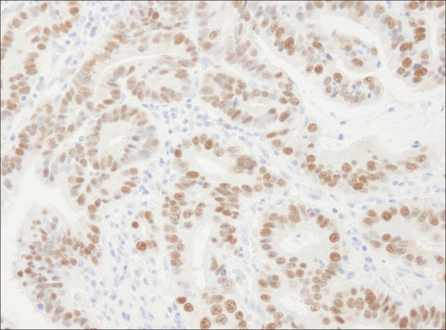
Cat. No. PLA0079: Detection of Human PCNA by Immunohistochemistry. Sample: FFPE section of Human stomach carcinoma. Antibody: Affinity purified Rabbit Anti-PCNA (
Cat. No. PLA0079) used at a dilution of 1:10,000 (0.1 μg/mL). Detection: DAB
 Immunofluorescence
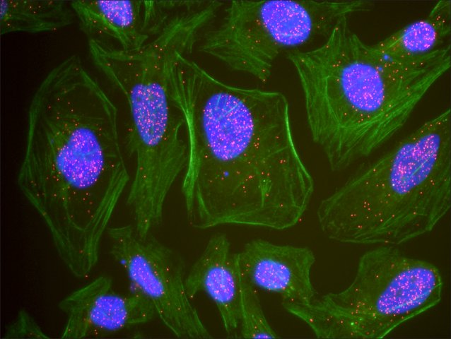
: This antibody is qualified for the Proximity Ligation Assay (PLA). The above image shows representative results for PLA using three color fluorescence, including DAPI stained nuclei (blue), phalloidin stained cytoplasmic F-actin (green), and PLA positive signal (red).
 Immunoblotting
Rabbit Anti-PCNA Antibody, Affinity Purified:
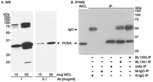
Cat. No. PLA0079: Detection of Human PCNA by Western Blot and Immunoprecipitaiton. Samples: A. Whole cell lysate (WCL) from A-431 cells. B. WCL (500 μg for IP; 30 μg for WCL lane) from MDA-MB-468 cells. Antibodies: A. Affinity purified Rabbit Anti-PCNA Antibody (
Cat. No. PLA0079) used at the indicated concentrations for WB. B. PCNA was immunoprecipitated using Antibodies recognizing different epitopes (including Cat. No.
PLA0080) or a Mouse monoclonal Antibody to PCNA (mAb). Control mock IP was performed using normal Mouse IgG (M-IgG) and normal Rabbit IgG (R-IgG). Immunoprecipitated PCNA was blotted using one of the alternate Antibodies (each at 1 μg/mL). Detection: Chemiluminescence with an exposure time of 2 minutes (A) or 5 seconds (B).
 Flow Cytometry
Rabbit Anti-PCNA Antibody, Affinity Purified:
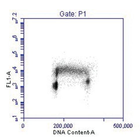
Cat. No. PLA0079: Flow Cytometric Detection of PCNA Versus DNA Content. Asynchronous Jurkat cells were fixed and permeabilized in a sequential treatment of FACS buffer (PBS, 0.5% Triton-X-100, 0.5 mM EDTA, 1% BSA) and 100% methanol. 1 × 10
6 cells were stained with 0.03 μg Anti-PCNA (
PLA0079). Secondary detection was performed with FITC conjugated Goat F(ab′)2 Anti-Rabbit Antibody, and DNA stained with PI.


