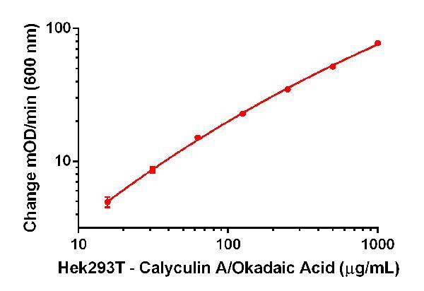
A dilution series of extract in Incubation Buffer in the working range of the assay. The extract was prepared from pellets of Hek293T cells treated with calyculin A + okadaic acid.
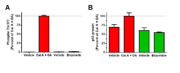
Cells were treated with calyculin A + okadaic acid (Cal A + OA, in red), etoposide (ab120227, in green) and corresponding drugs’ vehicles, as indicated. Diluted cell extracts were analyzed by this kit (A) and p53 protein ELISA (B), using ab117995. Extract of Hek 293T treated with calyculin A + okadaic acid was used for positive control sample standard curves. Relative levels interpolated from standard curves and expressed in percent of calyculin A + okadaic acid treated samples are shown.
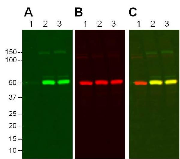
Hek293T cells were treated with vehicle (lane 1) or calyculin A + okadaic acid for 1 hour (lane 2) and 3 hours (lane 3). Cell extracts (40 µg/lane) were analyzed by Western blotting using the p53 pThr377 Detector Antibody of this kit and the p53 protein capture antibody of this kit. The image of p53 pT377 green IR signal is shown in A, the image of p53 protein red IR signal is shown in B, and an overlay of the images is shown in C.
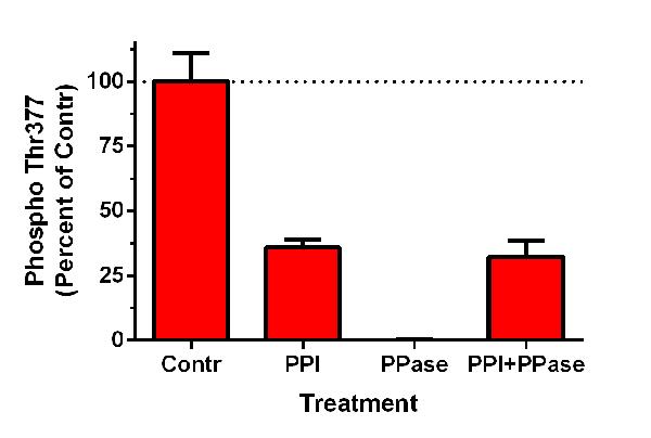
Extracts of Hek 293T cells (induced with calyculin A + okadaic acid) were treated with protein phosphatase inhibitor cocktail (PPI), protein phosphatase (PPase), protein phosphatase inhibitor cocktail and protein phosphatase (PPI+PPase) at 32 C for 30 minutes, or left untreated (Contr). Relative phospho Thr377 levels were determined using this kit
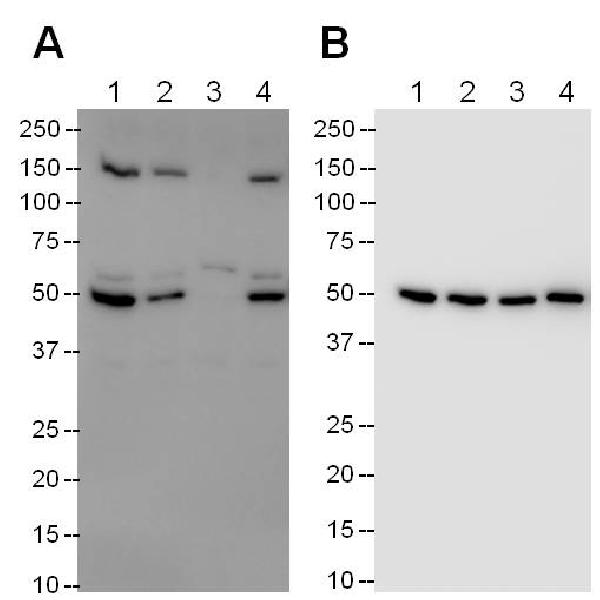
Extracts of Hek293T cells (induced with calyculin A + okadaic acid) were either treated with protein phosphatase inhibitor cocktail (PPI, lane 2), λ protein phosphatase (PPase, lane 3), PPI+PPase (lane 4) at 32 C for 30 minutes, or left untreated (lane 1). Samples (16 µg/lane) were analyzed by Western blotting using the p53 pThr377 Detector Antibody of this kit (A) or the p53 protein capture antibody of this kit (B).


