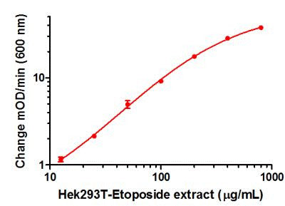
Example positive control sample standard curve. A dilution series of extract in Incubation Buffer in the working range of the assay. The extract was prepared from Hek293T cells treated with Etoposide by direct lysis (Protocol Booklet Section 7.2).

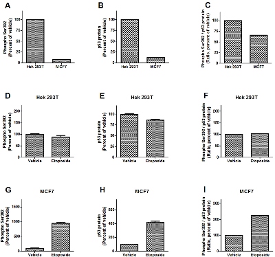
Example experimental analysis of drug treatment of Hek 293T and MCF7 cells. Cells were treated with Camptothecin, Etoposide or drug’s vehicle, as indicated. Diluted cell extracts (Hek293T to 200 µg/mL and MCF7 to 100 µg/mL) were analyzed by this kit (A, D, G) and p53 protein ELISA (B, E, H, using ab117995). Extract of Hek 293T treated with Etoposide was used for positive control sample standard curves. Relative levels interpolated from standard curves are shown. The levels of phosphorylated Ser392 normalized to total p53 protein levels were obtained as a ratio of phospho Ser392 and p53 protein levels (C, F, I).

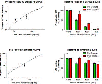
The p53 Phospho S392 ELISA specifically measures the phosphorylated Serine. In green: extracts of Hek 293T cells (induced with Etoposide) were treated with increasing concentrations of λ protein phosphatase or left untreated (Contr), and phospho Ser392 and total p53 protein levels were determined, respectively, using this kit and ab117995. In red: extracts of Hek 293T cells (induced with Etoposide) were immunocaptured to the plate, the immunocaptured materials were treated with increasing concentrations of λ protein phosphatase or left untreated (Contr), and phospho Ser392 and total p53 protein levels were determined using, respectively, this kit and ab117995. Dilutions of extracts of Hek 293T cells treated with Etoposide were used to construct the standard curves.

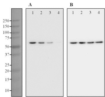
The detector antibody used in this kit specifically detects the phosphorylated p53 as determined by Western blotting. Extracts of Hek 293T cells (induced with Etoposide) were treated with increasing concentrations of λ protein phosphatase (lane 2, 400x diluted; lane 3, 100x diluted; lane 4, 25x diluted) or left untreated (lane 1). Samples were analyzed by Western blotting using the p53 Phospho S392 Detector Antibody (A). The membrane was re-probed to detect total p53 protein using the capture antibody of this kit (B).


