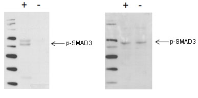
Using Western blot, SMAD3 phosphorylation at Ser423/425 is detected in TGF-treated C2C12 cells (+), compared with untreated C2C12 cells (-).
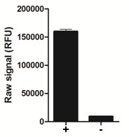
Using the p-SMAD3 assay kit, SMAD3 phosphorylation at Ser423/425 is detected in TGF-treated C2C12 cells (+) compared with untreated C2C12 cells.
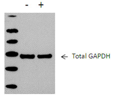
Using Western blot, cellular GAPDH is readily detected in A431 cellular lysates, in either untreated cells (-), or cells treated with EGF (+).
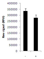
Using the GAPDH assay kit, cellular GAPDH is readily detected in A431 cellular lysates, in either untreated cells (-), or cells treated with EGF (+).
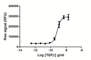
Raw264.7 cells were seeded at 80K cells/well in a 96 well tissue culture microplate overnight. The next day cells were serum starved for 60 minutes, then stimulated with various concentrations of TGFbeta for 60 min. The medium was removed from the wells, and cells were lysed with 120 µl/well of Lysis Mix, with shaking for 10 min. The lysates were transferred to a PhosphoTracer assay plate and assayed for phospho-SMAD3, using the standard protocol. Signal in the wells was determined using a plate reader.