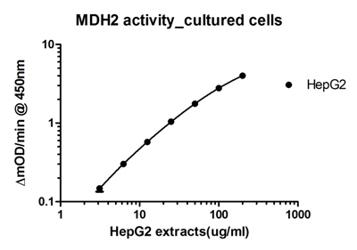
Figure 1. MDH2 activity measurements of serially diluted cultured HepG2 cell extracts.

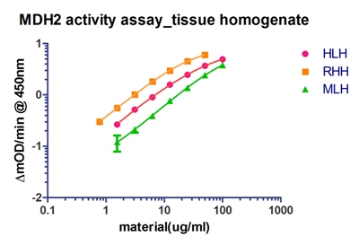
Figure 2. MDH2 activity measurements of serially diluted human liver homogenate, rat heart homogenate, and mouse liver homogenate.

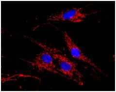
Figure 4. MDH2 antibody showing reactivity in a mitochondrial intracellular pattern with immunofluorescence microscopy.

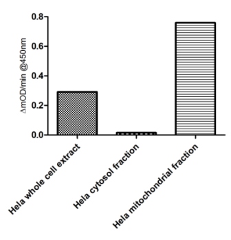
Figures 7. The isoform specificity of the malate activity measured by this kit is confirmed by measuring the MDH activity from different cell fractions. Activity was only detected from the mitochondrial fraction (MDH2), not the cytosol fraction (MDH1).


