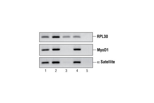
FIGURE 2. Chromatin immunoprecipitations were performed using digested chromatin from HeLa cells and either Histone H3 (D2B12) XP ® Rabbit mAb (ChIP Formulated) #4620 (lane 2), Rpb1 CTD (4H8) Mouse mAb #2629 (lane 3), Di-Methyl Histone H3 (Lys9) Antibody #9753 (lane 4), or Normal Rabbit IgG #2729 (lane 5). Purified DNA was analyzed by standard PCR methods using SimpleChIP ® Human RPL30 Exon 3 Primers #7014, SimpleChIP ® Human MyoD1 Exon 1 Primers #4490, and SimpleChIP ® Human α Satellite Repeat Primers #4486. PCR products were observed for each primer set in the input sample (lane 1) and various ChIP samples, but not in the Normal Rabbit IgG ChIP sample (lane 5).

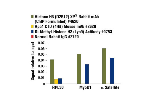
FIGURE 3. Chromatin immunoprecipitations were performed using digested chromatin from HeLa cells and the indicated ChIP-validated antibodies. Purified DNA was analyzed by quantitative real-time PCR, using SimpleChIP ® Human RPL30 Exon 3 Primers #7014 (control primer set), SimpleChIP ® Human MyoD1 Exon 1 Primers #4490, and SimpleChIP ® Human α Satellite Repeat Primers #4486. The amount of immunoprecipitated DNA in each sample is represented as signal relative to the total amount of input chromatin (equivalent to 1).

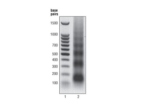
FIGURE 1. HeLa cells were formaldehyde-crosslinked and chromatin was prepared and digested as described in Section A of protocol. DNA was purified as described in Section B and 10 μl were separated by electrophoresis on a 1% agarose gel (lane 2) and stained with ethidium bromide. Lane 2 shows that the majority of chromatin was digested to 1 to 5 nucleosomes in length (150 to 900 bp).


