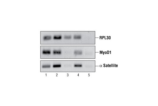
FIGURE 2. Chromatin immunoprecipitations were performed using digested chromatin from HeLa cells and either Histone H3 (D2B12) XP ® Rabbit mAb (ChIP Formulated) #4620 (lane 2), Rpb1 CTD (4H8) Mouse mAb #2629 (lane 3), Di-Methyl Histone H3 (Lys9) Antibody #9753 (lane 4) or Normal Rabbit IgG #2729 (lane 5). Purified DNA was analyzed by standard PCR methods using SimpleChIP ® Human RPL30 Exon 3 Primers #7014, SimpleChIP ® Human MyoD1 Exon 1 Primers #4490, and SimpleChIP ® Human α Satellite Repeat Primers #4486. PCR products were observed for each primer set in the input sample (lane 1) and various protein-specific immunoprecipitations but no PCR products were observed with immunoprecipitation using Normal Rabbit IgG #2729 (lane 5).

FIGURE 3. Chromatin immunoprecipitations were performed using digested chromatin from HeLa cells and the indicated ChIP-validated antibodies. Purified DNA was analyzed by quantitative real-time PCR, using SimpleChIP ® Human RPL30 Exon 3 Primers #7014 (control primer set), SimpleChIP ® Human MyoD1 Exon 1 Primers #4490, and SimpleChIP ® Human α Satellite Repeat Primers #4486. The amount of immunoprecipitated DNA in each sample is represented as signal relative to the total amount of input chromatin (equivalent to 1).

FIGURE 1. HeLa cells were formaldehyde-crosslinked and chromatin was prepared and digested as described in Section A of protocol. DNA was purified as described in Section B and 10 μl were separated by electrophoresis on a 1% agarose gel (lane 2) and stained with ethidium bromide. Lane 2 shows that the majority of chromatin was digested to 1 to 5 nucleosomes in length (150 to 900 bp).


