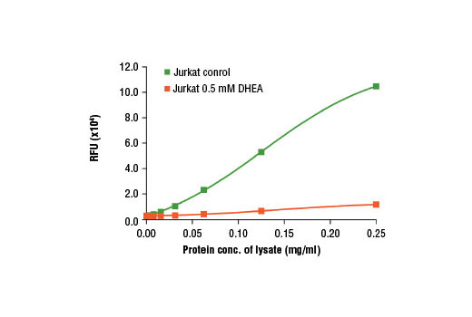
Figure 3. The relationship between the protein concentration of lysates from untreated and G6PD inhibitor DHEA (0.5 mM) treated Jurkat cells and relative fluorescence (RFU) is shown. The G6PD inhibitor DHEA can effectively inhibit this chain reaction as shown in this figure.

Figure 1. Schematic diagram of glucose-6-phosphate dehydrogenase (G6PD) assay. Glucose-6-phosphate (G6P) is oxidized by G6PD in the presence of NADP, which generates 6-phosphogluconolactone and NADPH. The generated NADPH is then amplified by the diaphorase-cycling system to produce highly fluorescent resorufin molecules.

Figure 2. Each assay component is individually omitted from the assay system and the resultant RFU is compared to that of a control test that contains all of the assay components.


