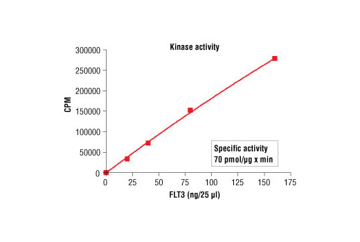
Figure 1. FLT3 kinase activity was measured in a radiometric assay using the following reaction conditions: 5 mM MOPS, pH 7.2, 2.5 mM β-glycerophosphate, 1 mM EGTA, 0.4 mM EDTA, 5 mM MgCl2, 0.05 mM DTT, 50 μM ATP, Substrate: MBP 200 ng/μL, and variable amounts of FLT3.

Figure 5. Staurosporine inhibition of FLT3 kinase activity: DELFIA® data generated using Phospho-Tyrosine mAb (P-Tyr-100) #9411 to detect phosphorylation of FLT3 substrate peptide (#1310) by FLT3 kinase. In a 50 µl reaction, 50 ng FLT3, 1.5 µM substrate peptide, 20 µM ATP and increasing amounts of staurosporine were used per reaction at room temperature for 30 minutes. (DELFIA® is a registered trademark of PerkinElmer, Inc.)

Figure 3. Dose dependence curve of FLT3 kinase activity: DELFIA® data generated using Phospho-Tyrosine mAb (P-Tyr-100) #9411 to detect phosphorylation of substrate peptide (#1310) by FLT3 kinase. In a 50 µl reaction, increasing amounts of FLT3 and 1.5 µM substrate peptide were used per reaction at room temperature for 30 minutes. (DELFIA® is a registered trademark of PerkinElmer, Inc.)

Figure 4. Peptide concentration dependence of FLT3 kinase activity: DELFIA® data generated using Phospho-Tyrosine mAb (P-Tyr-100) #9411 to detect phosphorylation of substrate peptide (#1310) by FLT3 kinase. In a 50 µl reaction, 50 ng of FLT3 and increasing concentrations of substrate peptide were used per reaction at room temperature for 30 minutes. (DELFIA® is a registered trademark of PerkinElmer, Inc.)

Figure 2. Time course of FLT3 kinase activity: DELFIA® data generated using Phospho-Tyrosine mAb (P-Tyr-100) #9411 to detect phosphorylation of FLT3 substrate peptide (#1310) by FLT3 kinase. In a 50 µl reaction, 50 ng FLT3 and 1.5 µM substrate peptide were used per reaction. (DELFIA® is a registered trademark of PerkinElmer, Inc.)


