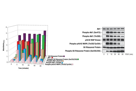
Figure 1. Treatment of NIH/3T3 cells with PDGF induces phosphorylation of Akt1 at Thr308 and Ser473, S6 Ribosomal Protein at Ser235/236 and p44/42 MAPK at Thr202/Tyr204 as detected by the PathScan ® Cell Growth Multi-Target Sandwich ELISA Kit #7239. While dynamic phosphorylation is observed throughout the time course, the level of total p44/42 MAPK, Akt1 and S6 ribosomal protein remains unchanged as demonstrated by sandwich ELISA and Western analysis. NIH/3T3 cells (80-90% confluent) were starved overnight and stimulated with PDGF (100 ng/mL) for 5, 10, 20, 40 and 80 minutes at 37ºC. Lysates were assayed at a protein concentration of 0.45 mg/mL. The absorbance readings at 450 nm are shown as a 3-dimensional representation in the left figure, while the corresponding Western blots are shown in the right figure. The antibodies used for the Western analyses include S6 Ribosomal Protein Rabbit mAb #2217, Phospho-S6 Ribosomal Protein (Ser235/236) Antibody #2211, Akt Antibody #9272, Phospho-Akt (Ser473) (193H12) Rabbit mAb #4058, Phospho-Akt (Thr308) Antibody #9275, Phospho-p44/42 MAPK (Thr202/Tyr204) (E10) Mouse mAb #9106, p44/42 MAP Kinase Antibody #9102.

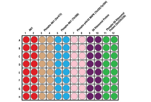
Figure 2. Schematic representation of a 96-well plate depicting the color-code of the reagents used to detect endogenous levels of Akt1 (red; 1 & 2), phospho-Akt1 (Ser473) (tan; 3 & 4), phospho-Akt1 (Thr308) (blue; 5 & 6), phospho-p44/42 MAPK (Thr202/Tyr204) (light pink; 7 & 8), S6 ribosomal protein (purple; 9 & 10) and phospho-S6 ribosomal protein (Ser235/236) (green; 11 & 12).

