Complex IV Human Protein Quantity Dipstick Assay Kit
| Name | Complex IV Human Protein Quantity Dipstick Assay Kit |
|---|---|
| Supplier | Abcam |
| Catalog | ab109877 |
| Prices | $343.00 |
| Sizes | 48 tests |
| Assay Type | Sandwich (quantitative) |
| Sample Type | Cell culture extracts, Tissue |
| Format | Reagents |
| Applications | ELISA |
| Species Reactivities | Bovine, Human |
| Gene | COX1 |
| Supplier Page | Shop |
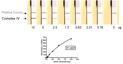
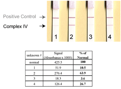
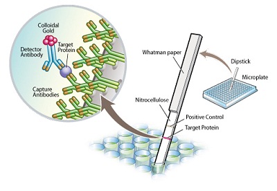
Product images
Product References
Dyclonine rescues frataxin deficiency in animal models and buccal cells of - Dyclonine rescues frataxin deficiency in animal models and buccal cells of
Sahdeo S, Scott BD, McMackin MZ, Jasoliya M, Brown B, Wulff H, Perlman SL, Pook MA, Cortopassi GA. Hum Mol Genet. 2014 Dec 20;23(25):6848-62.
Lateral-flow immunoassay for detecting drug-induced inhibition of mitochondrial - Lateral-flow immunoassay for detecting drug-induced inhibition of mitochondrial
Nadanaciva S, Willis JH, Barker ML, Gharaibeh D, Capaldi RA, Marusich MF, Will Y. J Immunol Methods. 2009 Mar 31;343(1):1-12.
Short hairpin RNA-mediated silencing of PRC (PGC-1-related coactivator) results - Short hairpin RNA-mediated silencing of PRC (PGC-1-related coactivator) results
Vercauteren K, Gleyzer N, Scarpulla RC. J Biol Chem. 2009 Jan 23;284(4):2307-19.
Mitochondrial oxidative phosphorylation protein levels in peripheral blood - Mitochondrial oxidative phosphorylation protein levels in peripheral blood
Shikuma CM, Gerschenson M, Chow D, Libutti DE, Willis JH, Murray J, Capaldi RA, Marusich M. AIDS Res Hum Retroviruses. 2008 Oct;24(10):1255-62.


