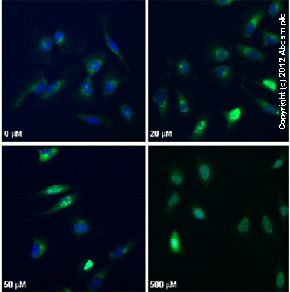
ab2893 staining γH2AX (phospho S139) in HeLa cells treated with camptothecin (ab120115), by ICC/IF. Increased nuclear expression of γH2AX (phospho S139) correlates with increased concentration of camptothecin, as described in literature.The cells were incubated at 37°C for 3h in media containing different concentrations of ab120115 (camptothecin) in DMSO, fixed with 4% formaldehyde for 10 minutes at room temperature and blocked with PBS containing 10% goat serum, 0.3 M glycine, 1% BSA and 0.1% tween for 2h at room temperature. Staining of the treated cells with ab2893 (10 µg/ml) was performed overnight at 4°C in PBS containing 1% BSA and 0.1% tween. A DyLight 488 goat anti-rabbit polyclonal antibody (ab96899) at 1/250 dilution was used as the secondary antibody. Nuclei were counterstained with DAPI and are shown in blue.

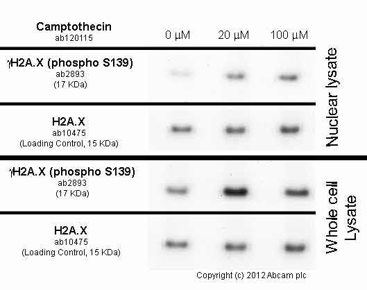
HeLa cells were incubated at 37°C for 3h with vehicle control (0 µM) and different concentrations of camptothecin (ab120115). Decreased expression of γH2A.X (phospho S139) in HeLa cells correlates with an increase camptothecin concentration, as described in literature.Whole cell lysates were prepared with RIPA buffer (containing protease inhibitors and sodium orthovanadate), 20µg of each were loaded on the gel and the WB was run under reducing conditions. After transfer the membrane was blocked for an hour using 5% BSA before being incubated with ab2893 at 1 µg/ml and ab10475 at 1 µg/ml overnight at 4°C. Antibody binding was detected using an anti-rabbit antibody conjugated to HRP (ab97051) at 1/10000 dilution and visualised using ECL development solution.

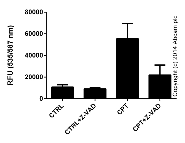
Functional assays: Caspase 3 (active) Red Staining Kit (ab65617) Caspase 3 activity in Jurkat cells (3 x10e5 cells) following 24 hour exposure to 2 uM Camptothecin (ab120115) with or without 50 μM caspase inhibitor Z-VAD(OMe)-FMK (ab120487). Background signal subtracted, duplicates; +/- SD.


