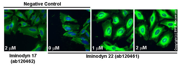
ab66705 staining PAI1 in HeLa cells treated with iminodyn-22™ (ab120461), by ICC/IF. Increase in PAI1 expression correlates with increased concentration of iminodyn-22™, as described in literature.The cells were incubated at 37°C for 48h in media containing different concentrations of ab120461 (Iminodyn-22™) in DMSO, fixed with 100% methanol for 5 minutes at -20°C and blocked with PBS containing 10% goat serum, 0.3 M glycine, 1% BSA and 0.1% tween for 2h at room temperature. Staining of the treated cells with ab66705 (5 µg/ml) was performed overnight at 4°C in PBS containing 1% BSA and 0.1% tween. A DyLight 488 goat anti-rabbit polyclonal antibody (ab96899) at 1/250 dilution was used as the secondary antibody. Nuclei were counterstained with DAPI and are shown in blue.

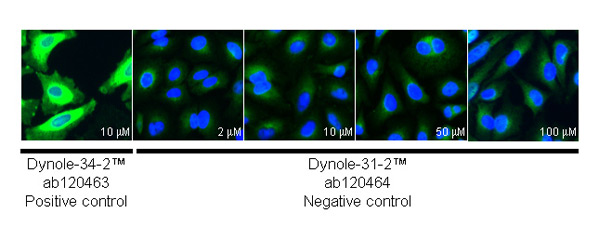
ab66705 staining PAI1 in HeLa cells treated with dynole-31-2™ (ab120464), by ICC/IF. No change in PAI1 expression with increased concentration of dynole-31-2™ (negative control for dynole 34-2™ (ab120463), as described in literature.The cells were incubated at 37°C for 6h in media containing different concentrations of ab120464 (dynole-31-2™) in DMSO, fixed with 100% methanol for 5 minutes at -20°C and blocked with PBS containing 10% goat serum, 0.3 M glycine, 1% BSA and 0.1% tween for 2h at room temperature. Staining of the treated cells with ab66705 (5 µg/ml) was performed overnight at 4°C in PBS containing 1% BSA and 0.1% tween. A DyLight 488 goat anti-rabbit polyclonal antibody (ab96899) at 1/250 dilution was used as the secondary antibody. Nuclei were counterstained with DAPI and are shown in blue.

ab66705 staining PAI1 in HeLa cells treated with OcTMAB™ (ab120467), by ICC/IF. Increase in PAI1 expression correlates with increased concentration of OcTMAB™, as described in literature.The cells were incubated at 37°C for 24h in media containing different concentrations of ab120467 (OcTMAB™) in DMSO, fixed with 100% methanol for 5 minutes at -20°C and blocked with PBS containing 10% goat serum, 0.3 M glycine, 1% BSA and 0.1% tween for 2h at room temperature. Staining of the treated cells with ab66705 (5 µg/ml) was performed overnight at 4°C in PBS containing 1% BSA and 0.1% tween. A DyLight 488 goat anti-rabbit polyclonal antibody (ab96899) at 1/250 dilution was used as the secondary antibody. Nuclei were counterstained with DAPI and are shown in blue.

ab66705 staining PAI1 in HeLa cells treated with dynole-34-2™; (ab120463), by ICC/IF. Increase in PAI1 expression correlates with increased concentration of dynole-34-2™, as described in literature.The cells were incubated at 37°C for 24h in media containing different concentrations of ab120463 (dynole-34-2™) in DMSO, fixed with 100% methanol for 5 minutes at -20°C and blocked with PBS containing 10% goat serum, 0.3 M glycine, 1% BSA and 0.1% tween for 2h at room temperature. Staining of the treated cells with ab66705 (5 µg/ml) was performed overnight at 4°C in PBS containing 1% BSA and 0.1% tween. A DyLight 488 goat anti-rabbit polyclonal antibody (ab96899) at 1/250 dilution was used as the secondary antibody. Nuclei were counterstained with DAPI and are shown in blue.

ab66705 staining PAI1 in HeLa cells treated with MiTMAB™ (ab120466), by ICC/IF. Increase in PAI1 expression correlates with increased concentration of MiTMAB™, as described in literature.The cells were incubated at 37°C for 24h in media containing different concentrations of ab120466 (MiTMAB™) in DMSO, fixed with 100% methanol for 5 minutes at -20°C and blocked with PBS containing 10% goat serum, 0.3 M glycine, 1% BSA and 0.1% tween for 2h at room temperature. Staining of the treated cells with ab66705 (5 µg/ml) was performed overnight at 4°C in PBS containing 1% BSA and 0.1% tween. A DyLight 488 goat anti-rabbit polyclonal antibody (ab96899) at 1/250 dilution was used as the secondary antibody. Nuclei were counterstained with DAPI and are shown in blue.