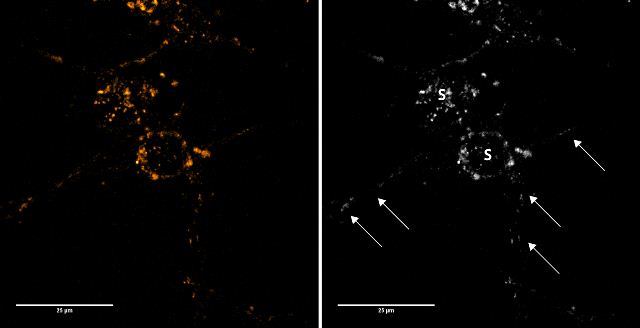
All images provided by Thorsten LauFigure 1: Two neuronal cells stained with 50 μM FFN102 on differentiation day 10. Shown is a sum projection of a confocal z-stack. Accumulation of FFN102 can be observed along the neurites (arrows) and the cell soma (S).

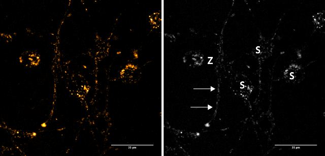
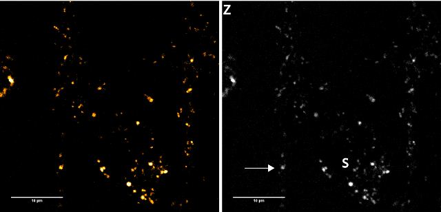
Figure 2a: Images in the first row show a group of neuronal cells stained with 50 μM FFN102 (sum projection of a confocal stack). FFN102 localizes to structures on the cell soma (S) as well as neurites (arrows). Z indicates the area zoomed in for an additional z-stack.

Figure 2b: Zoomed in area of “Z” from figure 2b. The arrow indicates a globular structure on a neurite. S indicates FFN102 positive structures on the cell soma.

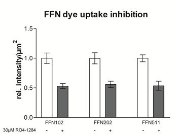
Figure 3: FFN102 dye uptake inhibition on addition of VMAT2 inhibitor RO4-1284


