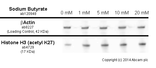
developed using the ECL techniquePerformed under reducing conditions.Exposure time : 10 seconds HeLa cells were incubated at 37°C for 6h with vehicle control (0 µM) and different concentrations of sodium butyrate (ab120948). Increased expression of histone H3 (acetyl K27) (ab4729) in HeLa cells correlates with an increase in sodium butyrate concentration, as described in literature.Whole cell lysates were prepared with RIPA buffer (containing protease inhibitors and sodium orthovanadate), 2.5µg of each were loaded on the gel and the WB was run under reducing conditions. After transfer the membrane was blocked for an hour using 5% BSA before being incubated with ab4927 at 1 µg/ml and ab8227 at 1 µg/ml overnight at 4°C. Antibody binding was detected using an anti-rabbit antibody conjugated to HRP (ab97051) at 1/10000 dilution and visualised using ECL development solution.

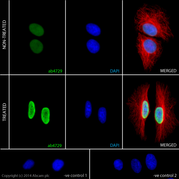
ab4729 staining Histone H3 (acetyl K27) in HeLa cells. The cells were incubated with 10mM Sodium butyrate (ab120948) for 6 hours (Treated) or solvent-only for control purposes (Non-treated). Cells were fixed with 100% methanol (5min) and then blocked in 1% BSA/10% normal goat serum/0.3M glycine in 0.1%PBS-Tween for 1h. The cells were then incubated with ab4729 at 0.5µg/ml and ab7291 at 1µg/ml overnight at +4°C, followed by a further incubation at room temperature for 1h with a anti-rabbit AlexaFluor®488 secondary antibody (ab150077) at 2 µg/ml (shown in green) and a goat anti-mouse AlexaFluor®594 (ab150120) at 2 µg/ml (shown in pseudo colour red). Nuclear DNA was labelled in blue with DAPI. Negative controls: 1– Rabbit primary and anti-mouse secondary antibody; 2 – Mouse primary antibody and anti-rabbit secondary antibody. Controls 1 and 2 indicate that there is no unspecific reaction between primary and secondary antibodies used.

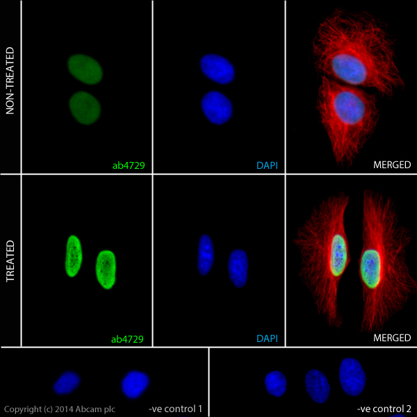
ab4729 staining Histone H3 (acetyl K27) in HeLa cells. The cells were incubated with 10mM Sodium butyrate (ab120948) for 6 hours (Treated) or solvent-only for control purposes (Non-treated). Cells were fixed with 100% methanol (5min) and then blocked in 1% BSA/10% normal goat serum/0.3M glycine in 0.1%PBS-Tween for 1h. The cells were then incubated with ab4729 at 0.5µg/ml and ab7291 at 1µg/ml overnight at +4°C, followed by a further incubation at room temperature for 1h with a anti-rabbit AlexaFluor®488 secondary antibody (ab150077) at 2 μg/ml (shown in green) and a goat anti-mouse AlexaFluor®594 (ab150120) at 2 μg/ml (shown in pseudo colour red). Nuclear DNA was labelled in blue with DAPI.Negative controls: 1– Rabbit primary and anti-mouse secondary antibody; 2 – Mouse primary antibody and anti-rabbit secondary antibody. Controls 1 and 2 indicate that there is no unspecific reaction between primary and secondary antibodies used.