Human M-CSF Receptor - Fc Chimera
| Name | Human M-CSF Receptor - Fc Chimera |
|---|---|
| Supplier | Abcam |
| Catalog | ab83682 |
| Prices | $315.00 |
| Sizes | 25 µg |
| Applications | FA SDS-PAGE |
| Species Reactivities | Human |
| Nature | Recombinant |
| Source | HEK 293 cells |
| Purity | > 95 % by SDS-PAGE. |
| Bioactivity | Activity: The ED50 of ab83682 is typically 0.2 - 0.3 ug/ml as measured by its ability to neutralize MCSF mediated proliferation of mNFS-60 cells. |
| SwissProt/Accession | P07333 |
| Gene | CSF1R |
| Residue | 20 to 512 |
| Sequence | Theoretical Sequence: IPVIEPSVPELVVKPGATVTLRCVGNGSVEWDGPPSPHWTLYSDGSSSIL STNNAT FQNTGTYRCTEPGDPLGGSAAIHLYVKDPARPWNVLAQEVVV FEDQDALLPCLLT DPVLEAGVSLVRVRGRPLMRHTNYSFSPWHGFTIH RAKFIQSQDYQCSALMGGRK VMSISIRLKVQKVIPGPPALTLVPAELV RIRGEAAQIVCSASSVDVNFDVFLQHNNTK LAIPQQSDFHNNRYQKVL TLNLDQVDFQHAGNYSCVASNVQGKHSTSMFFRVVES AYLNLSSEQNL IQEVTVGEGLNLKVMVEAYPGLQGFNWTYLGPFSDHQPEPKLAN ATTK DTYRHTFTLSLPRLKPSEAGRYSFLARNPGGWRALTFELTLRYPPEVSVI WT FINGSGTLLCAASGYPQPNVTWLQCSGHTDRCDEAQVLQVWDDPYP EVLSQEPF HKVTVQSLLTVETLEHNQTYECRAHNSVGSGSWAFIPISA GAHTHPPDEGSSNTKV DKKVEPKSCDKTHTCPPCPAPELLGGPSVFLF PPKPKDTLMISRTPEVTCVVVDVS HEDPEVKFNWYVDGVEVHNAKTKP REEQYNSTYRVVSVLTVLHQDWLNGKEYKC KVSNKALPAPIEKTISKA KGQPREPQVYTLPPSRDELTKNQVSLTCLVKGFYPSDIA VEWESNGQP ENNYKTTPPVLDSDGSFFLYSKLTVDKSRWQQGNVFSCSVMHEAL HNH YTQKSLSLSPGK |
| Supplier Page | Shop |
Product images
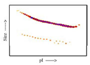 Densitometry of protein isoforms visualised by 2-DE. The triangle indicates the theoretical MW and pI of the protein.
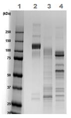 1D SDS-PAGE of ab83682 before and after treatment with glycosidases to remove oligosaccharides. Lane 1 MW markers; Lane 2 ab83682; Lane 3 ab83682 treated with PNGase F to remove potential N-linked glycans; Lane 4 ab83682 treated with a glycosidase cocktail to remove potential N- and O-linked glycans. Approximately 5 µg of protein was loaded per lane. Drop in MW after treatment with PNGase F indicates presence of N-linked glycans. A further drop in MW after treatment with the glycosidase cocktail indicates the presence of O-linked glycans. Additional bands in lane 3 and lane 4 are glycosidase enzymes.
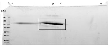 A sample of ab83682 without carrier protein was reduced and alkylated and focused on a 3-10 IPG strip then run on a 4-20% Tris-HCl 2D gel. Approximately 40 µg of protein was load; Gel was stained using Deep Purple™.