")
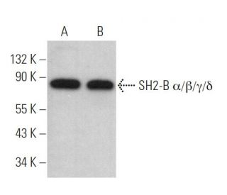
: sc-514142. Western blot analysis of SH2-B α/β/γ/δ expression in SJRH30 (A) and Sol8 (B) whole cell lysates. Detection reagent used: m-IgGκ BP-HRP: sc-516102.")
: sc-514142. Western blot analysis of SH2-B α/β/γ/δ expression in SJRH30 whole cell lysate.")
SH2-B α/β/γ/δ Antibody (C-11): sc-514142
- SH2-B α/β/γ/δ Antibody (C-11) is a mouse monoclonal IgG1 κ provided at 200 µg/ml
- specific for an epitope mapping between amino acids 2-24 at the N-terminus of SH2-B β of rat origin
- recommended for detection of SH2-B α, SH2-B β, SH2-B γ and SH2-B δ of mouse, rat and human origin by WB, IP, IF and ELISA
- Contact our Technical Service Department (or your local Distributor) for more information on how to receive a FREE 10 µg sample of SH2-B α/β/γ/δ (C-11): sc-514142.
- m-IgGκ BP-HRP is the preferred secondary detection reagent for SH2-B α/β/γ/δ Antibody (C-11) for WB applications. This reagent is now offered in a bundle with SH2-B α/β/γ/δ Antibody (C-11) (see ordering information below). For additional m-IgGκ BP conjugates see our complete list of Mouse IgG Binding Proteins.
QUICK LINKS
SEE ALSO...
SH2-B α/β/γ/δ Antibody (C-11) is a mouse monoclonal IgG1 kappa light chain antibody that detects SH2-B α/β/γ/δ in mouse, rat, and human samples through applications such as western blotting (WB), immunoprecipitation (IP), immunofluorescence (IF), and enzyme-linked immunosorbent assay (ELISA). Anti-SH2-B α/β/γ/δ antibody (C-11) is available in a non-conjugated form, allowing for versatile experimental setups. SH2-B, also known as SH2B1 or PSM, is a 756 amino acid protein that plays a crucial role in cellular signaling networks, particularly in regulating cell shape and movement, which is vital for tissue development and repair. SH2-B belongs to the APS family of adapter proteins, characterized by a pleckstrin homology (PH) domain, an SH2 domain, and a tyrosine phosphorylation site, which are essential for signal transduction. SH2-B is alternatively spliced to produce three distinct isoforms—SH2-B α, β, and γ—that share a common N-terminal sequence, including the PH and SH2 domains, along with multiple proline-rich motifs. These domains enable SH2-B to shuttle between the nucleus and cytoplasm, facilitating roles in various signaling pathways. SH2-B is predominantly expressed in skeletal muscle and ovary tissues, highlighting importance in muscle function and reproductive biology. Upon receptor kinase stimulation, SH2-B undergoes tyrosine phosphorylation, enhancing activity and interaction with other signaling molecules, thereby amplifying cellular responses to external stimuli.
Alexa Fluor® is a trademark of Molecular Probes Inc., OR., USA
LI-COR® and Odyssey® are registered trademarks of LI-COR Biosciences
SH2-B α/β/γ/δ Antibody (C-11) References:
- SH2-B is required for nerve growth factor-induced neuronal differentiation. | Rui, L., et al. 1999. J Biol Chem. 274: 10590-4. PMID: 10187854
- Identification of SH2-bbeta as a potent cytoplasmic activator of the tyrosine kinase Janus kinase 2. | Rui, L. and Carter-Su, C. 1999. Proc Natl Acad Sci U S A. 96: 7172-7. PMID: 10377387
- APS, an adapter protein with a PH and SH2 domain, is a substrate for the insulin receptor kinase. | Ahmed, Z., et al. 1999. Biochem J. 341 (Pt 3): 665-8. PMID: 10417330
- SH2-B, a membrane-associated adapter, is phosphorylated on multiple serines/threonines in response to nerve growth factor by kinases within the MEK/ERK cascade. | Rui, L., et al. 1999. J Biol Chem. 274: 26485-92. PMID: 10473609
- Differential binding to and regulation of JAK2 by the SH2 domain and N-terminal region of SH2-bbeta. | Rui, L., et al. 2000. Mol Cell Biol. 20: 3168-77. PMID: 10757801
- Regions of the JAK2 tyrosine kinase required for coupling to the growth hormone receptor. | Frank, SJ., et al. 1995. J Biol Chem. 270: 14776-85. PMID: 7540178
- Identification of SH2-Bbeta as a substrate of the tyrosine kinase JAK2 involved in growth hormone signaling. | Rui, L., et al. 1997. Mol Cell Biol. 17: 6633-44. PMID: 9343427
- Platelet-derived growth factor (PDGF) stimulates the association of SH2-Bbeta with PDGF receptor and phosphorylation of SH2-Bbeta. | Rui, L. and Carter-Su, C. 1998. J Biol Chem. 273: 21239-45. PMID: 9694882
Ordering Information
| Product Name | Catalog # | UNIT | Price | Qty | FAVORITES | |
SH2-B α/β/γ/δ Antibody (C-11) | sc-514142 | 200 µg/ml | $316.00 | |||
SH2-B α/β/γ/δ Antibody (C-11): m-IgGκ BP-HRP Bundle | sc-524620 | 200 µg Ab, 40 µg BP | $354.00 | |||
SH2-B α/β/γ/δ (C-11) Neutralizing Peptide | sc-514142 P | 100 µg/0.5 ml | $68.00 |