")
paxillin Antibody (C-1): sc-373880
- paxillin Antibody (C-1) is a mouse monoclonal IgG2a κ, cited in 5 publications, provided at 200 µg/ml
- specific for an epitope mapping between amino acids 17-47 near the N-terminus of paxillin of human origin
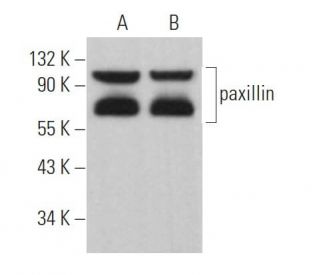
- recommended for detection of α,β, γ isoforms of paxillin of mouse, rat and human origin by WB, IP, IF and ELISA; also reactive with additional species, including and equine and canine
- See paxillin (B-2): sc-365379 for paxillin antibody conjugates, including AC, HRP, FITC, PE, Alexa Fluor® 488, 594, 647, 680 and 790.
- m-IgG2a BP-HRP and m-IgGκ BP-HRP are the preferred secondary detection reagents for paxillin Antibody (C-1) for WB applications. These reagents are now offered in bundles with paxillin Antibody (C-1) (see ordering information below).
QUICK LINKS
SEE ALSO...
paxillin Antibody (C-1) is a mouse monoclonal IgG2a kappa light chain antibody specific for an epitope mapping between amino acids 17-47 near the N-terminus of human paxillin. Paxillin, also known as PXN, is a focal adhesion phosphoprotein localized primarily to focal adhesions at the cytoplasmic face of the plasma membrane. The localization of paxillin is crucial because paxillin acts as a scaffold for assembling multiprotein complexes that mediate signal transduction between the extracellular matrix and the intracellular environment. Paxillin interacts with various proteins such as focal adhesion kinase (FAK), Src family kinases, vinculin, and integrins, playing a significant role in cell adhesion, migration, and proliferation. Phosphorylation of paxillin on specific tyrosine residues, notably Tyr118, modulates paxillin activity and interactions, impacting cellular processes like wound healing and cancer metastasis. During mitosis, paxillin levels decrease due to proteolytic downregulation, and paxillin undergoes alternative phosphorylation on serine residues, highlighting paxillin′s role in cell cycle regulation. anti-paxillin antibody (C-1) reacts with the α, β, and γ isoforms of paxillin from mouse, rat, human, equine, and canine origins in applications including western blotting (WB), immunoprecipitation (IP), immunofluorescence (IF), and enzyme-linked immunosorbent assay (ELISA). Understanding paxillin′s function and localization is important because alterations in paxillin expression or activity are associated with various diseases, making paxillin (C-1) antibody a valuable tool for researchers studying cell adhesion and signaling pathways.
Ordering Information
| Product Name | Catalog # | UNIT | Price | Qty | FAVORITES | |
paxillin Antibody (C-1) | sc-373880 | 200 µg/ml | $316.00 | |||
paxillin Antibody (C-1): m-IgGκ BP-HRP Bundle | sc-535324 | 200 µg Ab; 40 µg BP | $354.00 | |||
paxillin Antibody (C-1): m-IgG2a BP-HRP Bundle | sc-546221 | 200 µg Ab; 10 µg BP | $354.00 | |||
paxillin (C-1) Neutralizing Peptide | sc-373880 P | 100 µg/0.5 ml | $68.00 |