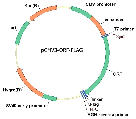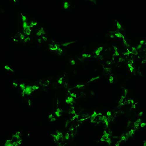
The plasmid was transfected into 293H adherent cells with Sinofection reagent (Cat# STF01). After 48 h, Immunofluorescence staining of cells. Cells were fixed with 4% PFA, permeabilzed with 0.3% Triton X-100 in PBS, blocked with 10% serum, and incubated with Mouse anti-Flag Tag monoclonal antibody (CST#8146S) at 37℃ 1 hour. Then cells were stained with Goat Anti-mouse IgG secondary antibody. The fluorescent signal is detected by fluorescence microscope. Each expression experiment has negative control.