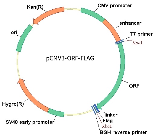
Human VEGFR2/KDR/Flk-1/CD309 Gene ORF cDNA clone expression plasmid, C-Flag tag
| Name | Human VEGFR2/KDR/Flk-1/CD309 Gene ORF cDNA clone expression plasmid, C-Flag tag |
|---|---|
| Supplier | Sino Biological |
| Catalog | HG10012-CF |
| Tag | Flag |
| Vector | pCMV3-C-FLAG |
| Species | Human |
| Gene | KDR |
| Description | Full length Clone DNA of Homo sapiens kinase insert domain receptor (a type I II receptor tyrosine kinase) with C terminal Flag tag. |
| Supplier Page | Shop |
Product images
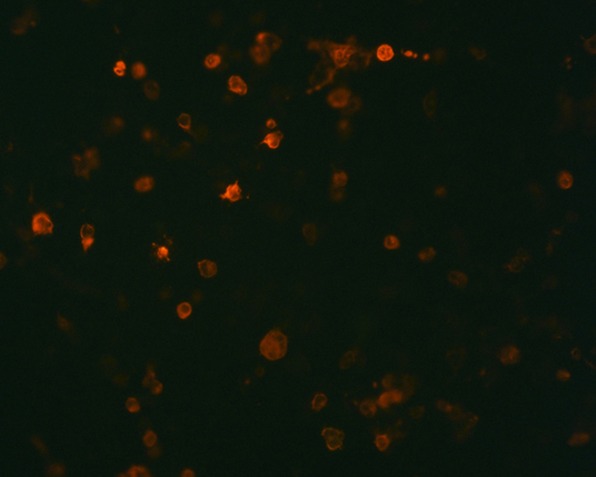 The plasmid was transfected into 293H adherent cells with Sinofection reagent (Cat# STF01). After 48 h, Immunofluorescence staining of cells. Cells were fixed with 4% PFA, permeabilzed with 0.3% Triton X-100 in PBS, blocked with 10% serum, and incubated with Mouse anti-Flag Tag monoclonal antibody (CST#8146S) at 37℃ 1 hour. Then cells were stained with Goat Anti-mouse IgG secondary antibody. The fluorescent signal is detected by fluorescence microscope. Each expression experiment has negative control.