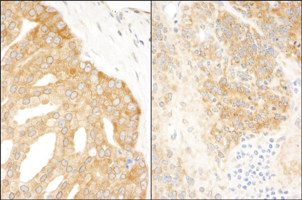
Immunohistochemistry (Formalin/PFA-fixed paraffin-embedded sections) analysis of human prostate carcinoma (left) and mouse teratoma (right) tissues labelling eIF3A with ab86146 at 1/200 (1µg/ml). Detection: DAB.
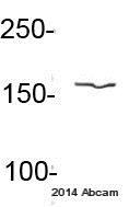
Anti-eIF3A antibody (ab86146) at 1/1000 dilution + Mouse pancreatic cancer whole cell lysate at 20 µgSecondaryIRDye® 680LT-conjugated donkey anti-mouse IgG polyclonal at 1/15000 dilutionPerformed under reducing conditions.
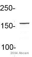
Anti-eIF3A antibody (ab86146) at 1/1000 dilution + Human U2OS osteosarcoma whole cell lysate at 20 µgSecondaryIRDye® 800CW conjugated donkey anti-rabbit polyclonal at 1/15000 dilutionPerformed under reducing conditions.
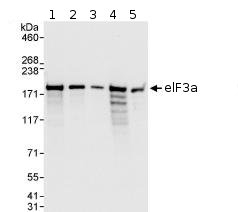
All lanes : Anti-eIF3A antibody (ab86146) at 0.04 µg/mlLane 1 : HeLa whole cell lysate at 50 µgLane 2 : HeLa whole cell lysate at 15 µgLane 3 : HeLa whole cell lysate at 5 µgLane 4 : 293T whole cell lysate at 50 µgLane 5 : NIH3T3 whole cell lysate at 50 µgdeveloped using the ECL technique
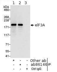
Detection of Human eIF3A by Immunoprecipitation, using ab86146 at 3µg/mg lysate. Image shows immunoprecipitated eIF3A detected with post IP WB, loading 20% of IP and using HeLa whole cell lysate at 1mg, with an antibody recognising an upstream epitope in lane 1, ab86146 at 1 µg/ml in lane 2 and control IgG in lane 3. Detection: Chemiluminescence with an exposure time of 3 seconds.


