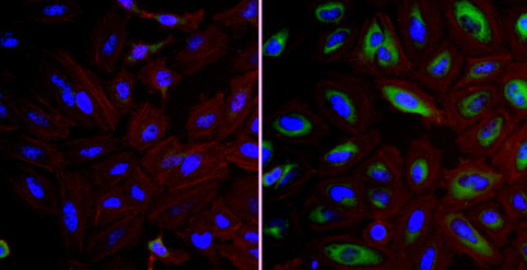
Immunocytochemistry/Immunofluorescence analysis of FKBP52 (green) in HeLa cells. Formalin fixed cells were permeabilized with 0.1% Triton X-100 in TBS for 10 minutes at room temperature and blocked with 1% BSA for 15 minutes at room temperature. Cells were probed without (left panel) or with (right panel) ab2926 (1:100) for at least 1 hour at room temperature, washed with PBS, and incubated with a DyLight 488-conjugated goat anti-rabbit IgG secondary antibody (1:400) for 30 minutes at room temperature. Nuclei (blue) were stained with Hoechst 33342 dye. Images were taken at 20X magnification.

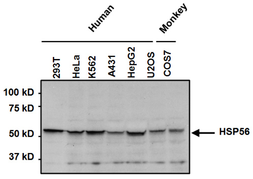
Western blot analysis of FKBP52 was performed by loading 50µg of the indicated whole cell lysates per well onto a 4-20% Tris-HCl polyacrylamide gel. Proteins were transferred to a PVDF membrane and blocked with 5% BSA/TBST for at least 1 hour. The membrane was incubated with ab2926 at a dilution of 1:1000 overnight at 4°C on a rocking platform, washed in TBS-0.1%Tween 20, and probed with a goat anti-rabbit IgG-HRP secondary antibody (1:20,000) for at least 1 hour. Chemiluminescent detection was performed.

Western blot of FKPB52 on human cell line S49 cytosol using ab2926.


