Anti-FOXP2 antibody
| Name | Anti-FOXP2 antibody |
|---|---|
| Supplier | Abcam |
| Catalog | ab16046 |
| Prices | $401.00 |
| Sizes | 100 µg |
| Host | Rabbit |
| Clonality | Polyclonal |
| Isotype | IgG |
| Applications | WB IHC-F IHC-F ICC/IF ICC/IF IHC-P |
| Species Reactivities | Mouse, Rat, Human |
| Antigen | Synthetic peptide conjugated to KLH derived from within residues 700 to the C-terminus of Human FOXP2 |
| Description | Rabbit Polyclonal |
| Gene | FOXP2 |
| Conjugate | Unconjugated |
| Supplier Page | Shop |
Product images
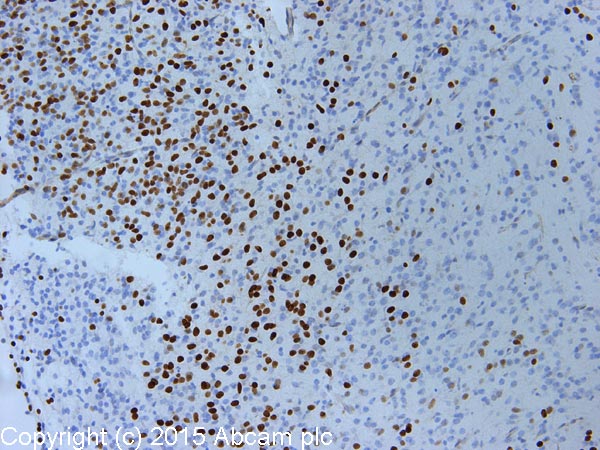 IHC image of FOXP2 staining in mouse e17 foetal brain formalin fixed paraffin embedded tissue section, performed on a Leica Bond™ system using the standard protocol B. The section was pre-treated using heat mediated antigen retrieval with sodium citrate buffer (pH6, epitope retrieval solution 1) for 20 mins. The section was then incubated with ab16046, 0.1µg/ml, for 15 mins at room temperature. A goat anti-rabbit biotinylated secondary antibody was used to detect the primary, and visualized using an HRP conjugated ABC system. DAB was used as the chromogen. The section was then counterstained with haematoxylin and mounted with DPX.For other IHC staining systems (automated and non-automated) customers should optimize variable parameters such as antigen retrieval conditions, primary antibody concentration and antibody incubation times.
 Mouse spinal cord was fixed in paraformaldehyde, blocked in 1% BSA for 30 minutes then incubated with ab16046 at 1/8000 dilution for 18 hours. This image was submitted as part of a review by Jeremy Dasen.
 This image is courtesy of an anonymous Abreviewab16046 at 1/1000 detecting FOXP2 from human 293T cell lysate (whole cell) (60ug/lane) by Western Blot. An HRP conjugated goat anti-rabbit IgG was used as the secondary and ECL was used as the detection method (1 minute exposure).See Abreview
 Image courtesy of Human Protein Atlasab16046 staining FOXP2 in human testis. Paraffin embedded human testis tissue was incubated with ab16046 (1/600 dilution) for 30 mins at room temperature. Antigen retrieval was performed by heat induction in citrate buffer pH 6. ab16046 was tested in a tissue microarray (TMA) containing a wide range of normal and cancer tissues as well as a cell microarray consisting of a range of commonly used, well characterised human cell lines. Further results for this antibody can be found at www.proteinatlas.org
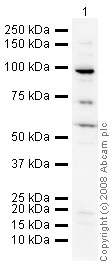 Anti-FOXP2 antibody (ab16046) at 1 µg/ml + HEK293 (Human embryonic kidney cell line) Whole Cell Lysate at 10 µgSecondaryGoat polyclonal to Rabbit IgG - H&L - Pre-Adsorbed (HRP) at 1/3000 dilutionPerformed under reducing conditions.Observed band size : 90 kDa (why is the actual band size different from the predicted?)Additional bands at : 56 kDa,70 kDa. We are unsure as to the identity of these extra bands.
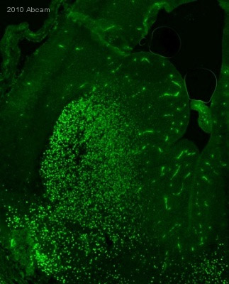 ab16046 staining FOXP2 in mouse brain tissue sections by IHC-Fr (Frozen sections). Tissue samples were fixed with paraformaldehyde, permeabilized by 0.4% Triton X and blocked with 10% serum for 1 hour at 22°C. The sample was incubated with primary antibody (1/8000) at 4°C for 16 hours. An Alexa Fluor®488-conjugated Goat polyclonal to mouse IgG (1/1000) was used as secondary antibody.See Abreview
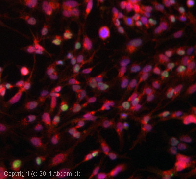 ICC/IF image of ab16046 stained HepG2 cells. The cells were 4% PFA fixed (10 min) and then incubated in 1%BSA / 10% normal goat serum / 0.3M glycine in 0.1% PBS-Tween for 1h to permeabilise the cells and block non-specific protein-protein interactions. The cells were then incubated with the antibody (ab16046, 1µg/ml) overnight at +4°C. The secondary antibody (green) was DyLight® 488 goat anti-rabbit IgG - H&L, pre-adsorbed (ab96899) used at a 1/250 dilution for 1h. Alexa Fluor® 594 WGA was used to label plasma membranes (red) at a 1/200 dilution for 1h. DAPI was used to stain the cell nuclei (blue) at a concentration of 1.43µM.
Product References
Integration of signals along orthogonal axes of the vertebrate neural tube - Integration of signals along orthogonal axes of the vertebrate neural tube
Sasai N, Kutejova E, Briscoe J. PLoS Biol. 2014 Jul 15;12(7):e1001907.
The scaffold protein Nde1 safeguards the brain genome during S phase of early - The scaffold protein Nde1 safeguards the brain genome during S phase of early
Houlihan SL, Feng Y. Elife. 2014 Sep 23;3:e03297.
Nhej1 Deficiency Causes Abnormal Development of the Cerebral Cortex. - Nhej1 Deficiency Causes Abnormal Development of the Cerebral Cortex.
El Waly B, Buhler E, Haddad MR, Villard L. Mol Neurobiol. 2014 Oct 7.
Ikaros promotes early-born neuronal fates in the cerebral cortex. - Ikaros promotes early-born neuronal fates in the cerebral cortex.
Alsio JM, Tarchini B, Cayouette M, Livesey FJ. Proc Natl Acad Sci U S A. 2013 Feb 19;110(8):E716-25. doi:
An epilepsy-related ARX polyalanine expansion modifies glutamatergic neurons - An epilepsy-related ARX polyalanine expansion modifies glutamatergic neurons
Beguin S, Crepel V, Aniksztejn L, Becq H, Pelosi B, Pallesi-Pocachard E, Bouamrane L, Pasqualetti M, Kitamura K, Cardoso C, Represa A. Cereb Cortex. 2013 Jun;23(6):1484-94.
Dicer is required for neural stem cell multipotency and lineage progression - Dicer is required for neural stem cell multipotency and lineage progression
Saurat N, Andersson T, Vasistha NA, Molnar Z, Livesey FJ. Neural Dev. 2013 Jul 29;8:14.
Cadm1-expressing synapses on Purkinje cell dendrites are involved in mouse - Cadm1-expressing synapses on Purkinje cell dendrites are involved in mouse
Fujita E, Tanabe Y, Imhof BA, Momoi MY, Momoi T. PLoS One. 2012;7(1):e30151.
Failed cytokinesis of neural progenitors in citron kinase-deficient rats leads to - Failed cytokinesis of neural progenitors in citron kinase-deficient rats leads to
Anastas SB, Mueller D, Semple-Rowland SL, Breunig JJ, Sarkisian MR. Cereb Cortex. 2011 Feb;21(2):338-44.
Robo1 regulates semaphorin signaling to guide the migration of cortical - Robo1 regulates semaphorin signaling to guide the migration of cortical
Hernandez-Miranda LR, Cariboni A, Faux C, Ruhrberg C, Cho JH, Cloutier JF, Eickholt BJ, Parnavelas JG, Andrews WD. J Neurosci. 2011 Apr 20;31(16):6174-87.
Abnormal development of the cerebral cortex and cerebellum in the setting of - Abnormal development of the cerebral cortex and cerebellum in the setting of
Coffinier C, Chang SY, Nobumori C, Tu Y, Farber EA, Toth JI, Fong LG, Young SG. Proc Natl Acad Sci U S A. 2010 Mar 16;107(11):5076-81. doi: