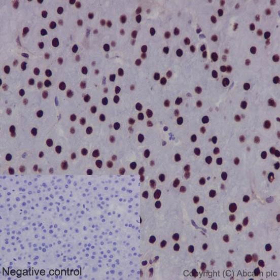
Immunohistochemical staining of paraffin embedded human hepatocellular carcinoma with purified ab126605 at a working dilution of 1 in 500. The secondary antibody used is a HRP polymer for rabbit IgG. The sample is counter-stained with hematoxylin. Antigen retrieval was perfomed using Tris-EDTA buffer, pH 9.0. PBS was used instead of the primary antibody as the negative control, and is shown in the inset.

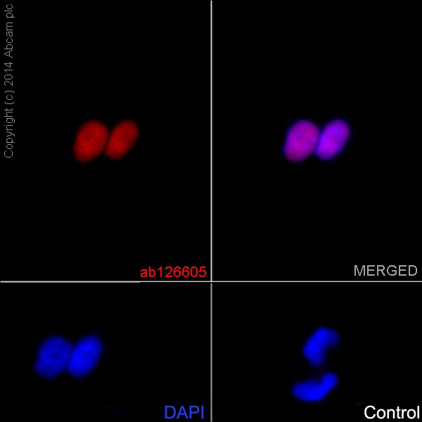
Immunofluorescence staining of BxPC-3 cells with purified ab126605 at a working dilution of 1 in 250, counter-stained with DAPI. The secondary antibody was Alexa Fluor® 555 goat anti rabbit, used at a dilution of 1 in 500. The cells were fixed in 4% PFA and permeabilized using 0.1% Triton X 100. The negative control is shown in bottom right hand panel - for the negative control, purified ab126605 was used at a dilution of 1/200 followed by an Alexa Fluor® 488 goat anti-mouse antibody at a dilution of 1/500.
![All lanes : Anti-FTO antibody [EPR6894] (ab126605) at 1/10000 dilution (purified)Lane 1 : Molt-4 cell lysateLane 2 : HEK293 cell lysateLane 3 : BxPC-3 cell lysateLane 4 : Caco-2 cell lysateLane 5 : SH-SY5Y cell lysateLysates/proteins at 20 µg per lane.SecondaryHRP goat anti-rabbit (H+L) at 1/1000 dilution](http://www.bioprodhub.com/system/product_images/ab_products/2/sub_2/21167_ab126605-241012-126605-WB.jpg)
All lanes : Anti-FTO antibody [EPR6894] (ab126605) at 1/10000 dilution (purified)Lane 1 : Molt-4 cell lysateLane 2 : HEK293 cell lysateLane 3 : BxPC-3 cell lysateLane 4 : Caco-2 cell lysateLane 5 : SH-SY5Y cell lysateLysates/proteins at 20 µg per lane.SecondaryHRP goat anti-rabbit (H+L) at 1/1000 dilution

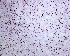
Immunohistochemical staining of FTO in paraffin embedded human adrenal gland tissue using unpurified ab126605 at a 1/50 dilution.

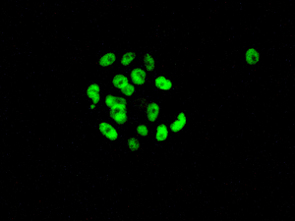
Unpurified ab126605 at a 1/50 dilution staining FTO in BxPC3 cells by immunofluorescence.
![All lanes : Anti-FTO antibody [EPR6894] (ab126605) at 1/1000 dilution (unpurified)Lane 1 : MOLT4 cell lysateLane 2 : 293T cell lysateLane 3 : SH SY5Y cell lysateLane 4 : BxPC3 cell lysateLane 5 : Caco 2 cell lysateLysates/proteins at 10 µg per lane.SecondaryGoat anti-Rabbit HRP at 1/2000 dilution](http://www.bioprodhub.com/system/product_images/ab_products/2/sub_2/21170_FTO-Primary-antibodies-ab126605-3.jpg)
All lanes : Anti-FTO antibody [EPR6894] (ab126605) at 1/1000 dilution (unpurified)Lane 1 : MOLT4 cell lysateLane 2 : 293T cell lysateLane 3 : SH SY5Y cell lysateLane 4 : BxPC3 cell lysateLane 5 : Caco 2 cell lysateLysates/proteins at 10 µg per lane.SecondaryGoat anti-Rabbit HRP at 1/2000 dilution


![All lanes : Anti-FTO antibody [EPR6894] (ab126605) at 1/10000 dilution (purified)Lane 1 : Molt-4 cell lysateLane 2 : HEK293 cell lysateLane 3 : BxPC-3 cell lysateLane 4 : Caco-2 cell lysateLane 5 : SH-SY5Y cell lysateLysates/proteins at 20 µg per lane.SecondaryHRP goat anti-rabbit (H+L) at 1/1000 dilution](http://www.bioprodhub.com/system/product_images/ab_products/2/sub_2/21167_ab126605-241012-126605-WB.jpg)


![All lanes : Anti-FTO antibody [EPR6894] (ab126605) at 1/1000 dilution (unpurified)Lane 1 : MOLT4 cell lysateLane 2 : 293T cell lysateLane 3 : SH SY5Y cell lysateLane 4 : BxPC3 cell lysateLane 5 : Caco 2 cell lysateLysates/proteins at 10 µg per lane.SecondaryGoat anti-Rabbit HRP at 1/2000 dilution](http://www.bioprodhub.com/system/product_images/ab_products/2/sub_2/21170_FTO-Primary-antibodies-ab126605-3.jpg)