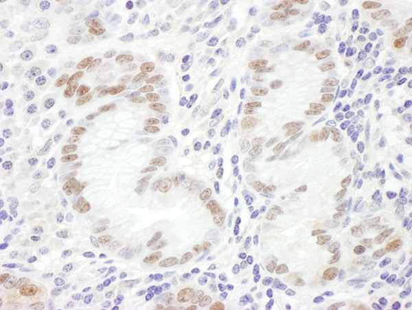
Immunohistochemistry (Formalin/PFA-fixed paraffin-embedded sections) analysis of human stomach linitis plastica tissue labelling GATA4 with ab124265 at 1/1000 (1µg/ml). Detection: DAB.

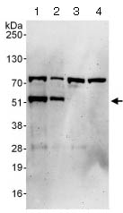
All lanes : Anti-GATA4 antibody (ab124265) at 0.1 µg/mlLane 1 : 293T whole cell lysate at 50 µg/mlLane 2 : 293T whole cell lysate at 15 µg/mlLane 3 : HeLa whole cell lysate at 50 µg/mlLane 4 : Jurkat whole cell lysate at 50 µg/mldeveloped using the ECL technique

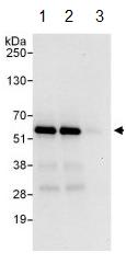
ab124265 at 1 µg/ml detecting GATA4 in 293T whole cell lysate by WB following IP. Lane 1: IP with an antibody which recognizes an upstream epitope of GATA4 Lane 2: ab124265 at 6 µg/mg of lysate Lane 3: Control IgG In each case, 1 mg of lysate was used for IP and 20% of the IP was loaded. Detection: Chemiluminescence with an exposure time of 10 seconds

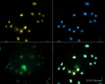
ab124265 staining GATA4 in human pluripotent stem cell derived cardiomyocytes by ICC/IF (Immunocytochemistry/immunofluorescence). Cells were fixed with paraformaldehyde, permeabilized with saponin and blocked with 5% serum for 15 minutes at room temperature. Samples were incubated with primary antibody (1/800) for 16 hours at 4°C. An Alexa Fluor® 568-conjugated goat anti-rabbit IgG monoclonal (1/1000) was used as the secondary antibody.Nuclear specific staining of GATA4 (yellow) in GFP positive cardiomyocytes (green), with GFP driven by a cardiac specific promoter.See Abreview


