Anti-Glutaminase antibody
| Name | Anti-Glutaminase antibody |
|---|---|
| Supplier | Abcam |
| Catalog | ab60709 |
| Prices | $400.00 |
| Sizes | 100 µg |
| Host | Mouse |
| Clonality | Monoclonal |
| Isotype | IgG2a |
| Applications | FC WB ELISA ICC/IF ICC/IF IP |
| Species Reactivities | Human |
| Antigen | Recombinant fragment with tag: EQRDYDSRTA LHVAAAEGHV EVVKFLLEAC KVNPFPKDRW NNTPMDEALH FGHHDVFKIL QEYQVQYTPQ GDSDNGKENQ TVHKNLDGLL , corresponding to amino acids 580-670 of Human Glutaminase Run BLAST with Run BLAST with |
| Description | Mouse Monoclonal |
| Gene | GLS |
| Conjugate | Unconjugated |
| Supplier Page | Shop |
Product images
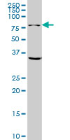 Anti-Glutaminase antibody (ab60709) + HeLa whole cell lysate at 50 µgSecondaryGoat Anti-Mouse IgG HRP at 1/2500 dilutiondeveloped using the ECL technique
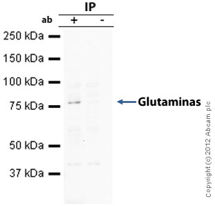 Glutaminase was immunoprecipitated using 0.5mg Hela whole cell extract, 10µg of Mouse monoclonal to Glutaminase (ab60709) and 50µl of protein G magnetic beads (+). No antibody was added to the control (-). The antibody was incubated under agitation with Protein G beads for 10min, Hela whole cell extract lysate diluted in RIPA buffer was added to each sample and incubated for a further 10min under agitation.Proteins were eluted by addition of 40µl SDS loading buffer and incubated for 10min at 70oC; 10µl of each sample was separated on a SDS PAGE gel, transferred to a nitrocellulose membrane, blocked with 5% BSA and probed with ab60709.Secondary: Goat polyclonal to mouse IgG light chain specific (HRP) at 1/5000 dilution.Band: 78kDa: Glutaminase.
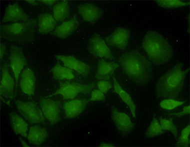 ab60709 at 10 ug/ml staining Glutaminase in human Hella cells by Immunocytochemistry/ Immunofluorescence.
![Overlay histogram showing HeLa cells stained with ab60709 (red line). The cells were fixed with 80% methanol (5 min) and then permeabilized with 0.1% PBS-Tween for 20 min. The cells were then incubated in 1x PBS / 10% normal goat serum / 0.3M glycine to block non-specific protein-protein interactions followed by the antibody (ab60709, 1µg/1x106 cells) for 30 min at 22°C. The secondary antibody used was DyLight® 488 goat anti-mouse IgG (H+L) (ab96879) at 1/500 dilution for 30 min at 22°C. Isotype control antibody (black line) was mouse IgG2a [ICIGG2A] (ab91361, 1µg/1x106 cells) used under the same conditions. Acquisition of >5,000 events was performed.](http://www.bioprodhub.com/system/product_images/ab_products/2/sub_2/25401_Glutaminase-Primary-antibodies-ab60709-5.jpg) Overlay histogram showing HeLa cells stained with ab60709 (red line). The cells were fixed with 80% methanol (5 min) and then permeabilized with 0.1% PBS-Tween for 20 min. The cells were then incubated in 1x PBS / 10% normal goat serum / 0.3M glycine to block non-specific protein-protein interactions followed by the antibody (ab60709, 1µg/1x106 cells) for 30 min at 22°C. The secondary antibody used was DyLight® 488 goat anti-mouse IgG (H+L) (ab96879) at 1/500 dilution for 30 min at 22°C. Isotype control antibody (black line) was mouse IgG2a [ICIGG2A] (ab91361, 1µg/1x106 cells) used under the same conditions. Acquisition of >5,000 events was performed.
Overlay histogram showing HeLa cells stained with ab60709 (red line). The cells were fixed with 80% methanol (5 min) and then permeabilized with 0.1% PBS-Tween for 20 min. The cells were then incubated in 1x PBS / 10% normal goat serum / 0.3M glycine to block non-specific protein-protein interactions followed by the antibody (ab60709, 1µg/1x106 cells) for 30 min at 22°C. The secondary antibody used was DyLight® 488 goat anti-mouse IgG (H+L) (ab96879) at 1/500 dilution for 30 min at 22°C. Isotype control antibody (black line) was mouse IgG2a [ICIGG2A] (ab91361, 1µg/1x106 cells) used under the same conditions. Acquisition of >5,000 events was performed.
Product References
Myc-induced AMPK-phospho p53 pathway activates Bak to sensitize mitochondrial - Myc-induced AMPK-phospho p53 pathway activates Bak to sensitize mitochondrial
Nieminen AI, Eskelinen VM, Haikala HM, Tervonen TA, Yan Y, Partanen JI, Klefstrom J. Proc Natl Acad Sci U S A. 2013 May 14;110(20):E1839-48. doi:
Glutamine synthetase is a genetic determinant of cell type-specific glutamine - Glutamine synthetase is a genetic determinant of cell type-specific glutamine
Kung HN, Marks JR, Chi JT. PLoS Genet. 2011 Aug;7(8):e1002229.
Glutamine metabolism is essential for human cytomegalovirus infection. - Glutamine metabolism is essential for human cytomegalovirus infection.
Chambers JW, Maguire TG, Alwine JC. J Virol. 2010 Feb;84(4):1867-73.