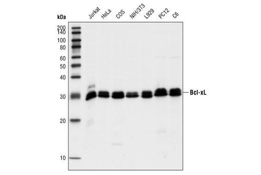
Western blot analysis of extracts from Jurkat and HeLa (human), COS (monkey), NIH/3T3 and L929 (mouse), and PC12 and C6 (rat) cells, using Bcl-xL (54H6) Rabbit mAb.

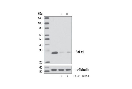
Western blot analysis of extracts from HeLa cells, transfected with 100 nM SignalSilence ® Control siRNA (Unconjugated) #6568 (-), SignalSilence ® BcL-xL siRNA I (+) or SignalSilence ® Bcl-xL siRNA II #6363 (+), using Bcl-xL (54H6) Rabbit mAb #2764 (upper) or α-Tubulin (11H10) Rabbit mAb #2125 (lower). The Bcl-xL (54H6) Rabbit mAb confirms silencing of Bcl-xL expression, while the α-Tubulin (11H10) Rabbit mAb is used as a loading control.

Immunoprecipitation of Bcl-xL from Jurkat cell extracts, using Bcl-xL (54H6) Rabbit mAb. Lane 1 is the lysate control, lane 2 is antibody alone and lane 3 is antibody plus lysate.

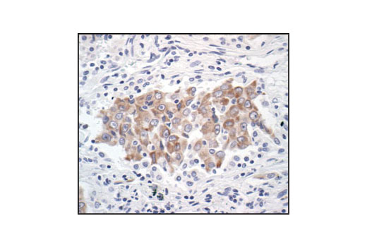
Immunohistochemical analysis of paraffin-embedded human lung carcinoma, using Bcl-xL (54H6) Rabbit mAb.

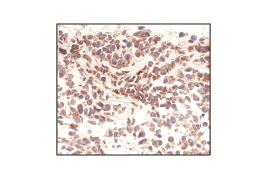
Immunohistochemical analysis of paraffin-embedded 4TI syngeneic mouse tumor, using Bcl-xL (54H6) Rabbit mAb # 2764.

Immunohistochemical analysis of paraffin-embedded human colon carcinoma, using Bcl-xL (54H6) Rabbit mAb in the presence of control peptide (left) or Bcl-xL Blocking Peptide #1225 (right).

Immunohistochemical analysis of paraffin-embedded human prostate carcinoma, showing cytoplasmic localization, using Bcl-xL (54H6) Rabbit mAb.

Immunohistochemical analysis of frozen H1650 xenograft, showing cytoplasmic localization using Bcl-xL (54H6) Rabbit mAb.

Confocal immunofluorescent analysis of HeLa cells using Bcl-xL (54H6) Rabbit mAb (green). Blue pseudocolor = DRAQ5® #4084 (fluorescent DNA dye).

Flow cytometric analysis of untreated Jurkat cells, using Bcl-xL (54H6) Rabbit mAb (blue) compared to a nonspecific negative control antibody (red).


