BiP (C50B12) Rabbit mAb
| Name | BiP (C50B12) Rabbit mAb |
|---|---|
| Supplier | Cell Signaling Technology |
| Catalog | 3177 |
| Prices | $99.00, $246.00 |
| Sizes | 20 µl (2 western blots), 100 µl (10 western blots) |
| Host | Rabbit |
| Clonality | Monoclonal |
| Isotype | IgG |
| Clone | C50B12 |
| Applications | WB IHC-P IHC-F FC |
| Species Reactivities | Human, Mouse |
| Antigen | BiP (C50B12) Rabbit mAb is produced by immunizing rabbits with a synthetic peptide corresponding to residues surrounding Gly584 of human BiP. |
| Description | Rabbit Monoclonal |
| Gene | HSPA5 |
| Supplier Page | Shop |
Product images
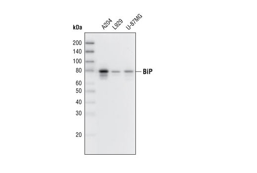 Western blot analysis of extracts from various cell lines using BiP (C50B12) Rabbit mAb.
 Immunohistochemical analysis of paraffin-embedded human glioblastoma using BiP (C50B12) Rabbit mAb.
 Immunohistochemical analysis of paraffin-embedded human colon carcinoma using BiP (C50B12) Rabbit mAb.
 Immunohistochemical analysis of paraffin-embedded human hepatocellular carcinoma using BiP (C50B12) Rabbit mAb.
 Immunohistochemical analysis of paraffin-embedded human breast carcinoma using BiP (C50B12) Rabbit mAb in the presence of control peptide (left) or BiP Blocking Peptide #1084 (right).
 Immunohistochemical analysis of frozen SKOV-3 xenograft using BiP (C50B12) Rabbit mAb.
 Flow cytometric analysis of A204 cells using BiP (C50B12) Rabbit mAb (blue) compared to concentration-matched Rabbit (DA1E) mAb IgG XP ® Isotype Control #3900 (red). Anti-rabbit IgG (H+L), F(ab') 2 Fragment (Alexa Fluor ® 488 Conjugate) #4412 was used as a secondary antibody.