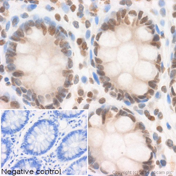
Anti-Histone H2B (acetyl ) antibody - ChIP Grade
| Name | Anti-Histone H2B (acetyl ) antibody - ChIP Grade |
|---|---|
| Supplier | Abcam |
| Catalog | ab1759 |
| Prices | $401.00 |
| Sizes | 100 µl |
| Host | Rabbit |
| Clonality | Polyclonal |
| Isotype | IgG |
| Applications | FC IHC-P IHC-F ChIP ELISA IP WB ICC/IF ICC/IF |
| Species Reactivities | Mouse, Rat, Human, Drosophila, Toxoplasma gondii |
| Antigen | Synthetic peptide corresponding to Histone H2B aa 9-20 |
| Description | Rabbit Polyclonal |
| Gene | HIST2H2BF |
| Conjugate | Unconjugated |
| Supplier Page | Shop |
Product images
Product References
Epigenetic regulations of immediate early genes expression involved in memory - Epigenetic regulations of immediate early genes expression involved in memory
Hendrickx A, Pierrot N, Tasiaux B, Schakman O, Kienlen-Campard P, De Smet C, Octave JN. PLoS One. 2014 Jun 11;9(6):e99467.
A novel activator of CBP/p300 acetyltransferases promotes neurogenesis and - A novel activator of CBP/p300 acetyltransferases promotes neurogenesis and
Chatterjee S, Mizar P, Cassel R, Neidl R, Selvi BR, Mohankrishna DV, Vedamurthy BM, Schneider A, Bousiges O, Mathis C, Cassel JC, Eswaramoorthy M, Kundu TK, Boutillier AL. J Neurosci. 2013 Jun 26;33(26):10698-712.
Drug inhibition of HDAC3 and epigenetic control of differentiation in Apicomplexa - Drug inhibition of HDAC3 and epigenetic control of differentiation in Apicomplexa
Bougdour A, Maubon D, Baldacci P, Ortet P, Bastien O, Bouillon A, Barale JC, Pelloux H, Menard R, Hakimi MA. J Exp Med. 2009 Apr 13;206(4):953-66.
.
ATF2 is required for amino acid-regulated transcription by orchestrating specific - ATF2 is required for amino acid-regulated transcription by orchestrating specific
Bruhat A, Cherasse Y, Maurin AC, Breitwieser W, Parry L, Deval C, Jones N, Jousse C, Fafournoux P. Nucleic Acids Res. 2007;35(4):1312-21. Epub 2007 Jan 31.
Myc-binding-site recognition in the human genome is determined by chromatin - Myc-binding-site recognition in the human genome is determined by chromatin
Guccione E, Martinato F, Finocchiaro G, Luzi L, Tizzoni L, Dall' Olio V, Zardo G, Nervi C, Bernard L, Amati B. Nat Cell Biol. 2006 Jul;8(7):764-70. Epub 2006 Jun 11.
Transcription-induced chromatin remodeling at the c-myc gene involves the local - Transcription-induced chromatin remodeling at the c-myc gene involves the local
Farris SD, Rubio ED, Moon JJ, Gombert WM, Nelson BH, Krumm A. J Biol Chem. 2005 Jul 1;280(26):25298-303. Epub 2005 May 6.


![Image from Bougdour A et al, J Exp Med 206:953-66 (2009), Fig S2.ab1759 used in Western Blot.FR235222 treatment specifically affects the acetylation status of histone H4 but not the acetylation levels of H3 and H2B. Extracellular T. gondii parasites (RH WT, R20D9 [TgHDAC3T99A], and M3135E11 [TgHDAC3T99I]) were treated with the indicated concentrations of FR235222 for 4 hours and lysed. 50µg of total cell lysates were analyzed by Immunoblotting with an anti-AcH4, anti-AcH2B (ab1759 at a 1/1000 dilution), anti-H3 and anti–alpha-tubulin antibodies.](http://www.bioprodhub.com/system/product_images/ab_products/2/sub_3/1270_Histone-H2B-Primary-antibodies-ab1759-1.jpg)
