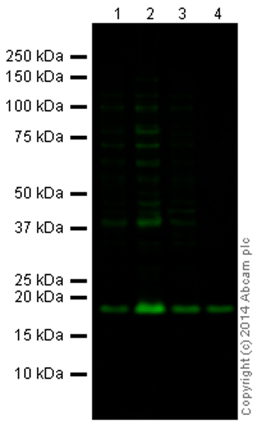
All lanes : Anti-Histone H3 (asymmetric di methyl R2) antibody - ChIP Grade (ab175007) at 1 µg/mlLane 1 : HeLa (Human epithelial carcinoma cell line) Whole Cell Lysate at 10 µgLane 2 : HeLa Nuclear Prep (0.5% Triton X-100 insoluble fraction) at 10 µgLane 3 : NIH 3T3 (Mouse embryonic fibroblast cell line) Whole Cell Lysate at 10 µgLane 4 : Calf Thymus Histone Preparation Nuclear Lysate at 0.5 µgSecondaryGoat Anti-Rabbit IgG H&L (Alexa Fluor® 790) (ab175781) at 1/10000 dilution

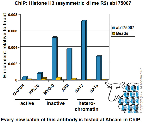
Chromatin was prepared from HeLa cells according to the Abcam X-ChIP protocol. Cells were fixed with formaldehyde for 10 minutes. The ChIP was performed with 25µg of chromatin, 2µg of ab175007 (blue), and 20µl of Protein A/G sepharose beads. No antibody was added to the beads control (yellow). The immunoprecipitated DNA was quantified by real time PCR (Taqman approach for active and inactive loci, Sybr green approach for heterochromatic loci). Primers and probes are located in the first kb of the transcribed region.

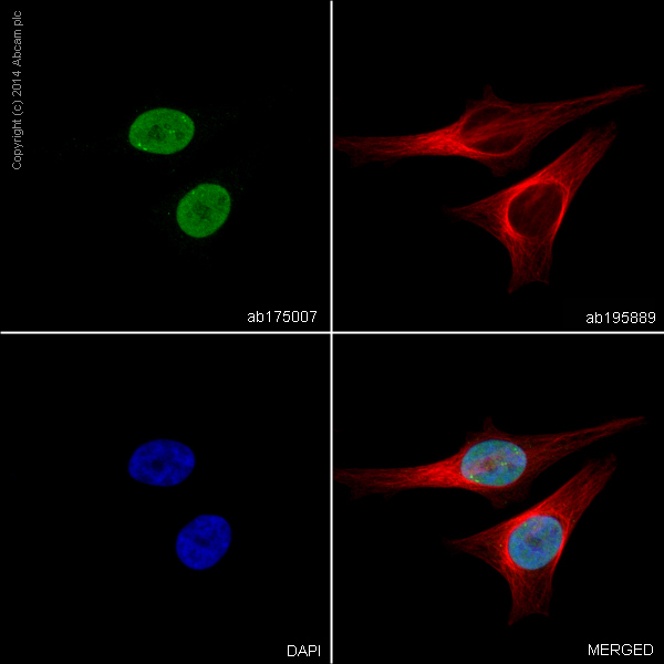
ab175007 staining Histone H3 (asymmetric di methyl R2) in HeLa cells. The cells were fixed with 4% PFA (10min), permabilised with 0.1%Triton (5min) and then blocked in 1% BSA/10% normal goat serum/0.3M glycine in 0.1%PBS-Tween for 1h. The cells were then incubated with ab175007 at 2μg/ml and ab195889 at 2µg/ml (shown in pseudo color red) overnight at +4°C, followed by a further incubation at room temperature for 1h with a goat anti-rabbit AlexaFluor®488 (ab150081) at 2 μg/ml (shown in green). Nuclear DNA was labelled in blue with DAPI.


