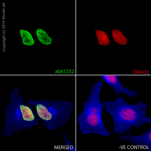
ab61252 staining Histone H3 in HeLa cells. The cells were fixed with 100% methanol (5min) and then blocked in 1% BSA/10% normal goat serum/0.3M glycine in 0.1%PBS-Tween for 1h. The cells were then incubated with ab61252 at 1μg/ml overnight at +4°C, followed by a further incubation at room temperature for 1h with a goat anti-rabbit AlexaFluor®488 secondary (ab150077) at 2 μg/ml (shown in green). AlexaFluor®350 WGA was used at a 1/200 dilution and incubated for 1h with the cells, to label plasma membranes (shown in blue). Nuclear DNA was labelled in red with 1.25 μM DRAQ5™ (ab108410), which was added to the secondary antibody mixture. A secondary only negative control is displayed, which indicates that the Histone H3 staining observed is due to primary antibody specificity and not to unspecific binding of the secondary antibody to the cells.

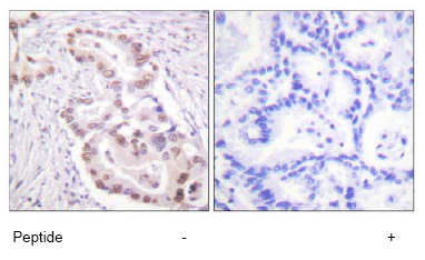
ab61252 at 1/50 - 1/100 dilution staining Histone H3 in human lung carcinoma by Immunohistochemistry, Paraffin-embedded tissue, in the absence or presence of the immunising peptide.

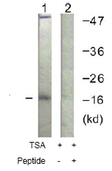
All lanes : Anti-Histone H3 antibody (ab61252) at 1/500 dilutionLane 1 : Jurkat cell extracts treated with TSA (400nM, 24hours)Lane 2 : Jurkat cell extracts treated with TSA (400nM, 24hours) with the immunising peptide


