Anti-HMGB1 antibody - ChIP Grade
| Name | Anti-HMGB1 antibody - ChIP Grade |
|---|---|
| Supplier | Abcam |
| Catalog | ab18256 |
| Prices | $404.00 |
| Sizes | 100 µg |
| Host | Rabbit |
| Clonality | Polyclonal |
| Isotype | IgG |
| Applications | IHC-F ICC/IF ICC/IF IHC-P ChIP IHC-F ICC/IF ELISA IP WB |
| Species Reactivities | Mouse, Rat, Human, Rabbit, Bovine |
| Antigen | Synthetic peptide conjugated to KLH derived from within residues 150 to the C-terminus of Human HMGB1 |
| Blocking Peptide | Human HMGB1 peptide |
| Description | Rabbit Polyclonal |
| Gene | HMGB1 |
| Conjugate | Unconjugated |
| Supplier Page | Shop |
Product images
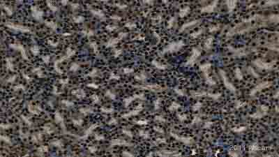
 Image courtesy of Human Protein AtlasParaffin embedded sections of human liver were incubated with ab18256 (1/1000 dilution) for 30 minutes at room temperature. Heat induced antigen retrieval was performed in citrate buffer pH 6.ab18256 was tested in a tissue microarray (TMA) containing a wide range of normal and cancer tissues as well as a cell microarray consisting of a range of commonly used, well characterised human cell lines. Further images can be found www.proteinatlas.org
 All lanes : Anti-HMGB1 antibody - ChIP Grade (ab18256) at 1/1000 dilutionLane 1 : Rat brain whole tissue lysate - infused with asf for 1 weekLane 2 : Rat brain whole tissue lysate - infused with LPS for 1 weekLane 3 : Rat brain whole tissue lysate - infused with acsf for 8 weeksLane 4 : Rat brain whole tissue lysate - infused with LPS for 8 weeksLane 5 : Rat brain whole tissue lysate - infused with LPS for 4 weeksLane 6 : Rat brain whole tissue lysate -infused with LPS for 4 weeks, after 2 weeks of LPS infusion were treated with neramexane for the next 2 weeksLane 7 : Rat brain whole tissue lysate - infused with LPS for 4 weeks, after 2 weeks of LPS infusion were treated with memantine for the next 2 weeks.Lysates/proteins at 40 µg per lane.SecondaryBiotinylated Goat anti-rabbit IgGdeveloped using the ECL techniquePerformed under reducing conditions.
 All lanes : Anti-HMGB1 antibody - ChIP Grade (ab18256) at 1 µg/mlLane 1 : NIH 3T3 (Mouse embryonic fibroblast cell line) Whole Cell Lysate (ab7179)Lane 2 : MEF1 (Mouse embryonic fibroblast cell line) Whole Cell LysateLane 3 : PC12 (Rat adrenal pheochromocytoma cell line) Whole Cell Lysate Lysates/proteins at 10 µg per lane.SecondaryIRDye 680 Conjugated Goat Anti-Rabbit IgG (H+L) at 1/10000 dilutionPerformed under reducing conditions.
 All lanes : Anti-HMGB1 antibody - ChIP Grade (ab18256) at 1 µg/mlLane 1 : HeLa (Human epithelial carcinoma cell line) Whole Cell Lysate Lane 2 : Jurkat (Human T cell lymphoblast-like cell line) Whole Cell Lysate (ab7899)Lane 3 : A431 (Human epithelial carcinoma cell line) Whole Cell Lysate (ab7909)Lane 4 : HEK293 (Human embryonic kidney cell line) Whole Cell Lysate (ab7902)Lysates/proteins at 10 µg per lane.SecondaryIRDye 680 Conjugated Goat Anti-Rabbit IgG (H+L) at 1/10000 dilutionPerformed under reducing conditions.
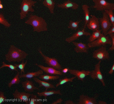
 ICC/IF image of ab18256 stained HeLa cells. The cells were 4% PFA fixed (10 min) and then incubated in 1%BSA / 10% normal goat serum / 0.3M glycine in 0.1% PBS-Tween for 1h to permeabilise the cells and block non-specific protein-protein interactions. The cells were then incubated with the antibody (ab18256, 1µg/ml) overnight at +4°C. The secondary antibody (green) was Alexa Fluor® 488 goat anti-rabbit IgG (H+L) used at a 1/1000 dilution for 1h. Alexa Fluor® 594 WGA was used to label plasma membranes (red) at a 1/200 dilution for 1h. DAPI was used to stain the cell nuclei (blue) at a concentration of 1.43µM. This antibody also gave a positive result in 4% PFA fixed (10 min) Hek293, HepG2 and MCF7 cells at 1µg/ml.
 ab18256 staining HMGB1 in Human stomach tissue sections by IHC-Fr (Immunohistochemistry - Frozen sections). Tissue samples were fixed with acetone and blocked with 5% serum for 1 hour at 25°C. Samples were incubated with primary antibody 1/500 in blocking buffer for 1 hour at 25°C. An undiluted HRP-conjugated Goat polyclonal to rabbit IgG was used as secondary antibody.See Abreview
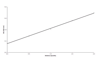 ab18256 used in Direct ELISA in NIH 3T3 murine fibroblasts. Primary antibody used at a 1/1000 dilution for 16 hours at 4°C. The secondary antibody is an AP-conjugated Goat anti-rabbit used at a 1/1000 dilution. A blocking step was performed using 5% BSA for 1 hour.See Abreview
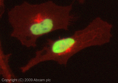
 ICC/IF image of ab18256 stained Hela cells. The cells were 4% PFA fixed (10 min) and then incubated in 1%BSA / 10% normal goat serum / 0.3M glycine in 0.1% PBS-Tween for 1h to permeabilise the cells and block non-specific protein-protein interactions. The cells were then incubated with the antibody (ab18256, 5µg/ml) overnight at +4°C. The secondary antibody (green) was DyLight® 488 goat anti-rabbit IgG - HandL, pre-adsorbed (ab96899) used at a 1/250 dilution for 1h. Alexa Fluor® 594 WGA was used to label plasma membranes (red) at a 1/200 dilution for 1h. DAPI was used to stain the cell nuclei (blue) at a concentration of 1.43µM.
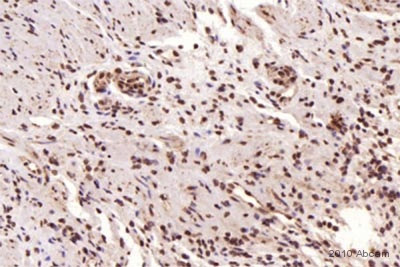
 ab18256 staining HMGB1 in murine kidney tissue by Immunohistochemistry (Formalin/PFA-fixed paraffin-embedded sections).Tissue was fixed in formaldehyde and a heat mediated antigen retrieval step was performed using citrate EDTA buffer pH 6.2. Samples were then blocked, then incubated with ab18256 at a 1/1000 dilution for 1 hour. The secondary used was a goat anti-rabbit IgG conjugated to HRP at a 1/1000 dilution.See Abreview
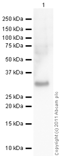 Anti-HMGB1 antibody - ChIP Grade (ab18256) at 1 µg/ml + Human HMGB1 full length protein (ab56525) at 0.01 µgSecondaryGoat Anti-Rabbit IgG H&L (HRP) preadsorbed (ab97080) at 1/5000 dilutiondeveloped using the ECL techniquePerformed under reducing conditions.Exposure time : 2 minutes
Product References
HMGB1 in severe soft tissue infections caused by Streptococcus pyogenes. - HMGB1 in severe soft tissue infections caused by Streptococcus pyogenes.
Johansson L, Snall J, Sendi P, Linner A, Thulin P, Linder A, Treutiger CJ, Norrby-Teglund A. Front Cell Infect Microbiol. 2014 Jan 30;4:4.
Cellular mechanisms of high mobility group 1 (HMGB-1) protein action in the - Cellular mechanisms of high mobility group 1 (HMGB-1) protein action in the
Santos AR, Dvoriantchikova G, Li Y, Mohammad G, Abu El-Asrar AM, Wen R, Ivanov D. PLoS One. 2014 Jan 31;9(1):e87574.
JAK/STAT1 signaling promotes HMGB1 hyperacetylation and nuclear translocation. - JAK/STAT1 signaling promotes HMGB1 hyperacetylation and nuclear translocation.
Lu B, Antoine DJ, Kwan K, Lundback P, Wahamaa H, Schierbeck H, Robinson M, Van Zoelen MA, Yang H, Li J, Erlandsson-Harris H, Chavan SS, Wang H, Andersson U, Tracey KJ. Proc Natl Acad Sci U S A. 2014 Feb 25;111(8):3068-73. doi:
Activation of the NLRP1b inflammasome independently of ASC-mediated caspase-1 - Activation of the NLRP1b inflammasome independently of ASC-mediated caspase-1
Van Opdenbosch N, Gurung P, Vande Walle L, Fossoul A, Kanneganti TD, Lamkanfi M. Nat Commun. 2014;5:3209.
Modeling Mycobacterium tuberculosis early granuloma formation in experimental - Modeling Mycobacterium tuberculosis early granuloma formation in experimental
Parasa VR, Rahman MJ, Ngyuen Hoang AT, Svensson M, Brighenti S, Lerm M. Dis Model Mech. 2014 Feb;7(2):281-8.
Release of neuronal HMGB1 by ethanol through decreased HDAC activity activates - Release of neuronal HMGB1 by ethanol through decreased HDAC activity activates
Zou JY, Crews FT. PLoS One. 2014 Feb 14;9(2):e87915.
HMGB1 localization during experimental periodontitis. - HMGB1 localization during experimental periodontitis.
Nogueira AV, de Souza JA, de Molon RS, Pereira Eda S, de Aquino SG, Giannobile WV, Cirelli JA. Mediators Inflamm. 2014;2014:816320.
Protective effect of glycyrrhizin, a direct HMGB1 inhibitor, on focal cerebral - Protective effect of glycyrrhizin, a direct HMGB1 inhibitor, on focal cerebral
Gong G, Xiang L, Yuan L, Hu L, Wu W, Cai L, Yin L, Dong H. PLoS One. 2014 Mar 4;9(3):e89450.
Curcumin: a double hit on malignant mesothelioma. - Curcumin: a double hit on malignant mesothelioma.
Miller JM, Thompson JK, MacPherson MB, Beuschel SL, Westbom CM, Sayan M, Shukla A. Cancer Prev Res (Phila). 2014 Mar;7(3):330-40. doi:
HMGB1-promoted and TLR2/4-dependent NK cell maturation and activation take part - HMGB1-promoted and TLR2/4-dependent NK cell maturation and activation take part
Qiu Y, Yang J, Wang W, Zhao W, Peng F, Xiang Y, Chen G, Chen T, Chai C, Zheng S, Watkins DJ, Feng J. PLoS Pathog. 2014 Mar 20;10(3):e1004011.