![Anti-HNF-4-alpha antibody [EPR16885] (ab181604) at 1/1000 dilution + Full length mouse HNF-4-gamma recombinant protein at 10 µgSecondaryGoat Anti-Rabbit IgG, (H+L),Peroxidase conjugated at 1/1000 dilution](http://www.bioprodhub.com/system/product_images/ab_products/2/sub_3/3140_ab181604-242418-wb-1.jpg)
Anti-HNF-4-alpha antibody [EPR16885] (ab181604) at 1/1000 dilution + Full length mouse HNF-4-gamma recombinant protein at 10 µgSecondaryGoat Anti-Rabbit IgG, (H+L),Peroxidase conjugated at 1/1000 dilution
![Anti-HNF-4-alpha antibody [EPR16885] (ab181604) at 1/10000 dilution + HepG2 (Human liver hepatocellular carcinoma) whole cell lysate at 10 µgSecondaryGoat Anti-Rabbit IgG, (H+L),Peroxidase conjugated at 1/1000 dilution](http://www.bioprodhub.com/system/product_images/ab_products/2/sub_3/3141_ab181604-242417-wb-2.jpg)
Anti-HNF-4-alpha antibody [EPR16885] (ab181604) at 1/10000 dilution + HepG2 (Human liver hepatocellular carcinoma) whole cell lysate at 10 µgSecondaryGoat Anti-Rabbit IgG, (H+L),Peroxidase conjugated at 1/1000 dilution
![Anti-HNF-4-alpha antibody [EPR16885] (ab181604) at 1/1000 dilution + SW480 (Human colon adenocarcinoma cell line) whole cell lysate at 10 µgSecondaryGoat Anti-Rabbit IgG, (H+L),Peroxidase conjugated at 1/1000 dilution](http://www.bioprodhub.com/system/product_images/ab_products/2/sub_3/3142_ab181604-242416-wb-3.jpg)
Anti-HNF-4-alpha antibody [EPR16885] (ab181604) at 1/1000 dilution + SW480 (Human colon adenocarcinoma cell line) whole cell lysate at 10 µgSecondaryGoat Anti-Rabbit IgG, (H+L),Peroxidase conjugated at 1/1000 dilution
![All lanes : Anti-HNF-4-alpha antibody [EPR16885] (ab181604) at 1/1000 dilutionLane 1 : Human fetal liver lysateLane 2 : Human colon lysateLane 3 : Human fetal kidney lysateLysates/proteins at 10 µg per lane.SecondaryAnti-Rabbit IgG (HRP), specific to the non-reduced form of IgG at 1/1000 dilution](http://www.bioprodhub.com/system/product_images/ab_products/2/sub_3/3143_ab181604-242415-wb-4.jpg)
All lanes : Anti-HNF-4-alpha antibody [EPR16885] (ab181604) at 1/1000 dilutionLane 1 : Human fetal liver lysateLane 2 : Human colon lysateLane 3 : Human fetal kidney lysateLysates/proteins at 10 µg per lane.SecondaryAnti-Rabbit IgG (HRP), specific to the non-reduced form of IgG at 1/1000 dilution
![All lanes : Anti-HNF-4-alpha antibody [EPR16885] (ab181604) at 1/1000 dilutionLane 1 : Mouse liver lysateLane 2 : Rat liver lysateLysates/proteins at 10 µg per lane.SecondaryGoat Anti-Rabbit IgG, (H+L),Peroxidase conjugated at 1/1000 dilution](http://www.bioprodhub.com/system/product_images/ab_products/2/sub_3/3144_ab181604-242414-wb-5.jpg)
All lanes : Anti-HNF-4-alpha antibody [EPR16885] (ab181604) at 1/1000 dilutionLane 1 : Mouse liver lysateLane 2 : Rat liver lysateLysates/proteins at 10 µg per lane.SecondaryGoat Anti-Rabbit IgG, (H+L),Peroxidase conjugated at 1/1000 dilution
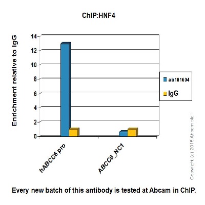
Chromatin was prepared from HepG2 (Human liver hepatocellular carcinoma) cells according to the Abcam X-ChIP protocol. Cells were fixed with formaldehyde for 10 minutes. The ChIP was performed with 25µg of chromatin, 2µg of ab181604 (blue), and 20µl of Anti rabbit IgG sepharose beads. 2μg of rabbit normal IgG was added to the beads control (yellow). The immunoprecipitated DNA was quantified by real time PCR (Sybr green approach).ABCC6_NC1 is negative control
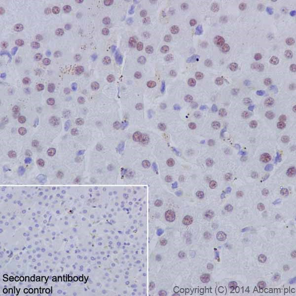
Immunohistochemical analysis of paraffin-embedded Human liver tissue labeling HNF-4-alpha with ab181604 at 1/2000 dilution, followed by Goat Anti-Rabbit IgG H&L (HRP) (ab97051) secondary antibody at 1/500 dilution. Nucleus staining on Human liver is observed. Counter stained with Hematoxylin.Secondary antibody only control: Used PBS instead of primary antibody, secondary antibody is Goat Anti-Rabbit IgG H&L (HRP) (ab97051) at 1/500 dilution.
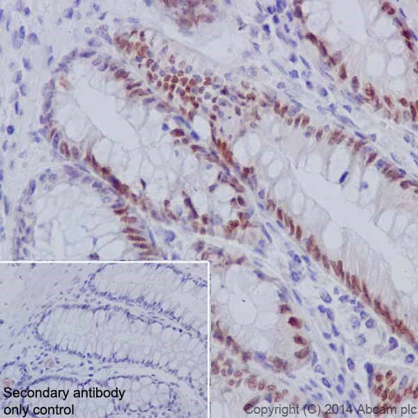
Immunohistochemical analysis of paraffin-embedded Human colon tissue labeling HNF-4-alpha with ab181604 at 1/2000 dilution, followed by Goat Anti-Rabbit IgG H&L (HRP) (ab97051) secondary antibody at 1/500 dilution. Nuclear staining on epithelial cells of Human colon is observed. Counter stained with Hematoxylin.Secondary antibody only control: Used PBS instead of primary antibody, secondary antibody is Goat Anti-Rabbit IgG H&L (HRP) (ab97051) at 1/500 dilution.
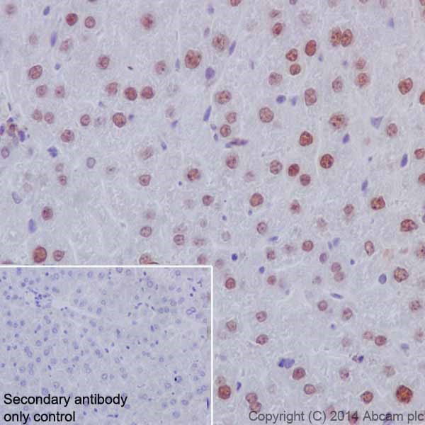
Immunohistochemical analysis of paraffin-embedded Mouse liver tissue labeling HNF-4-alpha with ab181604 at 1/2000 dilution, followed by Goat Anti-Rabbit IgG H&L (HRP) (ab97051) secondary antibody at 1/500 dilution. Nuclear staining on mouse liver is observed. Counter stained with Hematoxylin.Secondary antibody only control: Used PBS instead of primary antibody, secondary antibody is Goat Anti-Rabbit IgG H&L (HRP) (ab97051) at 1/500 dilution.
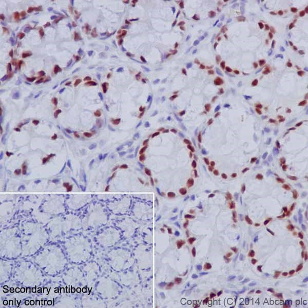
Immunohistochemical analysis of paraffin-embedded Rat colon tissue labeling HNF-4-alpha with ab181604 at 1/2000 dilution, followed by Goat Anti-Rabbit IgG H&L (HRP) (ab97051) secondary antibody at 1/500 dilution. Nuclear staining on epithelial cells of rat colon is observed. Counter stained with Hematoxylin.Secondary antibody only control: Used PBS instead of primary antibody, secondary antibody is Goat Anti-Rabbit IgG H&L (HRP) (ab97051) at 1/500 dilution.
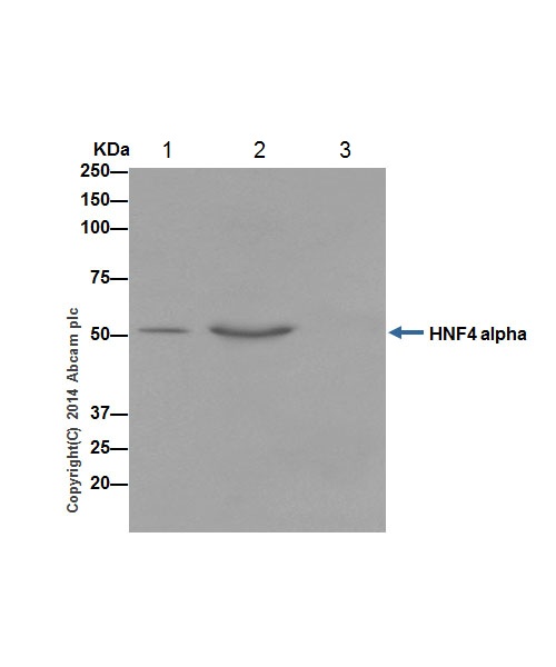
HNF-4 alpha was immunoprecipitated from 1mg of HepG2 (Human liver hepatocellular carcinoma) whole cell extract with ab181604 at 1/70 dilution. Western blot was performed from the immunoprecipitate using ab181604 at 1/5000 dilution. Anti-Rabbit IgG (HRP), specific to the non-reduced form of IgG, was used as secondary antibody at 1/1500 dilution.Lane 1: HepG2 whole cell extract 10 µg (Input).Lane 2: ab181604 IP in HepG2 whole cell extract.Lane 3: Rabbit monoclonal IgG (ab172730) instead of ab181604 in HepG2 whole cell extract.Blocking and dilution buffer and concentration: 5% NFDM/TBST.
![Anti-HNF-4-alpha antibody [EPR16885] (ab181604) at 1/1000 dilution + Full length mouse HNF-4-gamma recombinant protein at 10 µgSecondaryGoat Anti-Rabbit IgG, (H+L),Peroxidase conjugated at 1/1000 dilution](http://www.bioprodhub.com/system/product_images/ab_products/2/sub_3/3140_ab181604-242418-wb-1.jpg)
![Anti-HNF-4-alpha antibody [EPR16885] (ab181604) at 1/10000 dilution + HepG2 (Human liver hepatocellular carcinoma) whole cell lysate at 10 µgSecondaryGoat Anti-Rabbit IgG, (H+L),Peroxidase conjugated at 1/1000 dilution](http://www.bioprodhub.com/system/product_images/ab_products/2/sub_3/3141_ab181604-242417-wb-2.jpg)
![Anti-HNF-4-alpha antibody [EPR16885] (ab181604) at 1/1000 dilution + SW480 (Human colon adenocarcinoma cell line) whole cell lysate at 10 µgSecondaryGoat Anti-Rabbit IgG, (H+L),Peroxidase conjugated at 1/1000 dilution](http://www.bioprodhub.com/system/product_images/ab_products/2/sub_3/3142_ab181604-242416-wb-3.jpg)
![All lanes : Anti-HNF-4-alpha antibody [EPR16885] (ab181604) at 1/1000 dilutionLane 1 : Human fetal liver lysateLane 2 : Human colon lysateLane 3 : Human fetal kidney lysateLysates/proteins at 10 µg per lane.SecondaryAnti-Rabbit IgG (HRP), specific to the non-reduced form of IgG at 1/1000 dilution](http://www.bioprodhub.com/system/product_images/ab_products/2/sub_3/3143_ab181604-242415-wb-4.jpg)
![All lanes : Anti-HNF-4-alpha antibody [EPR16885] (ab181604) at 1/1000 dilutionLane 1 : Mouse liver lysateLane 2 : Rat liver lysateLysates/proteins at 10 µg per lane.SecondaryGoat Anti-Rabbit IgG, (H+L),Peroxidase conjugated at 1/1000 dilution](http://www.bioprodhub.com/system/product_images/ab_products/2/sub_3/3144_ab181604-242414-wb-5.jpg)


