![All lanes : Anti-htrA1 antibody [EPR14605(2)] (ab199529) at 1/5000 dilutionLane 1 : HepG2 (Human liver hepatocellular carcinoma) cell lysateLane 2 : MCF7 (Human breast adenocarcinoma cell line) cell lysateLysates/proteins at 10 µg per lane.SecondaryGoat Anti-Rabbit IgG, (H+L), Peroxidase conjugated at 1/1000 dilution](http://www.bioprodhub.com/system/product_images/ab_products/2/sub_3/5596_ab199529-241839-ab199529WB1.jpg)
All lanes : Anti-htrA1 antibody [EPR14605(2)] (ab199529) at 1/5000 dilutionLane 1 : HepG2 (Human liver hepatocellular carcinoma) cell lysateLane 2 : MCF7 (Human breast adenocarcinoma cell line) cell lysateLysates/proteins at 10 µg per lane.SecondaryGoat Anti-Rabbit IgG, (H+L), Peroxidase conjugated at 1/1000 dilution
![Anti-htrA1 antibody [EPR14605(2)] (ab199529) at 1/1000 dilution + Human fetal liver tissue lysate at 10 µgSecondaryAnti-Rabbit IgG (HRP), specific to the non-reduced form of IgG at 1/1000 dilution](http://www.bioprodhub.com/system/product_images/ab_products/2/sub_3/5597_ab199529-241844-ab199529WB2.jpg)
Anti-htrA1 antibody [EPR14605(2)] (ab199529) at 1/1000 dilution + Human fetal liver tissue lysate at 10 µgSecondaryAnti-Rabbit IgG (HRP), specific to the non-reduced form of IgG at 1/1000 dilution
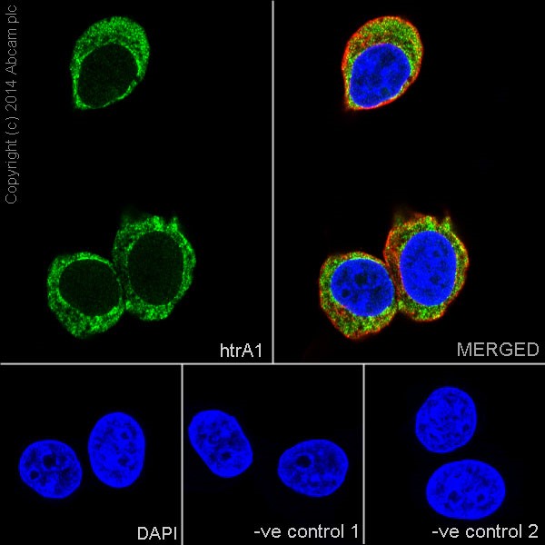
Immunofluorescent analysis of 4% paraformaldehyde-fixed, 0.1% Triton X-100 permeabilized MCF7 (Human breast adenocarcinoma cell line) cells labeling htrA1 with ab199529 at 1/1200 dilution, followed by Goat anti-rabbit IgG (Alexa Fluor® 488) (ab150077) secondary antibody at 1/500 (green). Cytoplasm staining on MCF7cell line is observed. The nuclear counterstain is DAPI (blue). Tubulin is detected with ab7291 (anti-Tubulin mouse mAb) at 1/1000 and ab150120 (AlexaFluor®594 Goat anti-Mouse secondary) at 1/500 dilution (red).The negative controls are as follows:--ve control 1: - ab199529 at 1/1200 dilution followed by ab150120 (AlexaFluor®594 Goat anti-Mouse secondary) at 1/500 dilution.-ve control 2: - ab7291 (anti-Tubulin mouse mAb) at 1/1000 dilution followed by ab150077 (Alexa Fluor®488 Goat Anti-Rabbit IgG H&L) at 1/500 dilution.
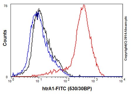
Flow cytometric analysis of 2% paraformaldehyde-fixed HepG2 (Human liver hepatocellular carcinoma) cells labeling htrA1 with ab199529 at 1/150 dilution (red), compared with a rabbit monoclonal IgG isotype control (ab172730, black) and an unlabelled control (cells without incubation with primary antibody and secondary antibody (blue). Goat anti rabbit IgG (FITC) at 1/150 dilution was used as the secondary antibody.
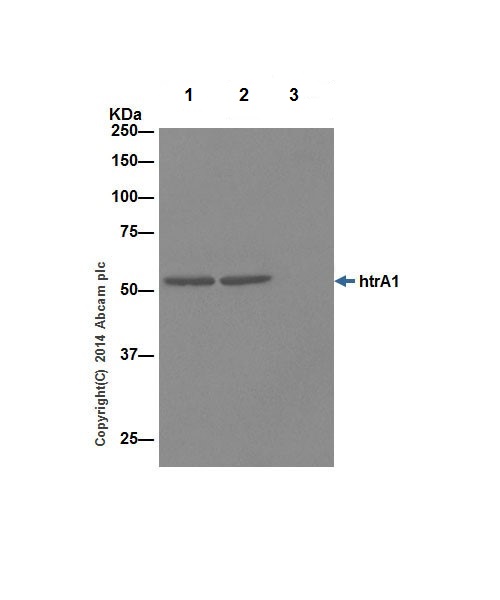
htrA1 was immunoprecipitated from 1mg of HepG2 (Human liver hepatocellular carcinoma) whole cell extract with ab199529 at 1/40 dilution. Western blot was performed using ab199529 at 1/1000 dilution. Anti-Rabbit IgG (HRP), specific to the non-reduced form of IgG, was used as secondary antibody at 1/1500 dilution.Lane 1: HepG2 whole cell extract 10 µg (Input).Lane 2: ab199529 IP in HepG2 whole cell extract.Lane 3: Rabbit monoclonal IgG (ab172730) instead of ab199529 in HepG2 whole cell extract.Blocking and dilution buffer and concentration: 5% NFDM/TBST.