Anti-Iba1 antibody
| Name | Anti-Iba1 antibody |
|---|---|
| Supplier | Abcam |
| Catalog | ab5076 |
| Prices | $400.00 |
| Sizes | 100 µg |
| Host | Goat |
| Clonality | Polyclonal |
| Isotype | IgG |
| Applications | IHC-F IHC-F IHC-P WB IHC-F ICC/IF ICC/IF ICC/IF |
| Species Reactivities | Rat, Rabbit, Guinea Pig, Bovine, Human, Pig, Marmoset, Mouse, Monkey |
| Antigen | Synthetic peptide: C-TGPPAKKAISELP , corresponding to amino acids 135-147 of Human Iba1(Peptide available as ab23067 |
| Description | Goat Polyclonal |
| Gene | AIF1 |
| Conjugate | Unconjugated |
| Supplier Page | Shop |
Product images
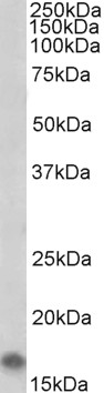 Anti-Iba1 antibody (ab5076) at 2 µg/ml + Rat Brain lysate at 35 µg
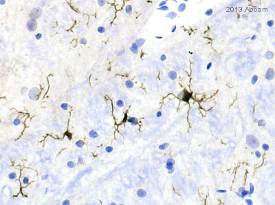
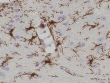
 Immunohistochemical analysis of paraffin embedded Mouse Brain tissue labeling Iba1 with ab5076 at 2 ug/ml. Tissue underwent antigen retrieval in steam with Tris/EDTA buffer (pH 9.0). The HRP-staining procedure was used for detection.
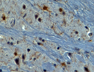
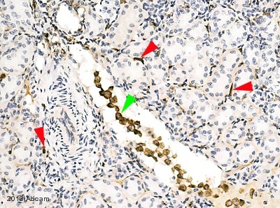
 IHC-P image of Iba1 staining on rat brain sections using ab5076 (1:1000). The sections were deparaffinized and subjected to heat mediated antigen retrieval using citric acid. The sections were blocked using 1% BSA for 10 mins at 21°C. ab5076 was diluted 1:1000 and incubted with the sections for 2 hours at 21°C. The secondary antibody used was Rabbit polyclonal to anti sheep/goat IgG conjugated to biotin (1:200)See Abreview
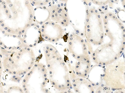 IHC-P image of Iba1 staining on cat kidney sections using ab5076 (1:1000). The sections were deparaffinized and subjected to heat mediated antigen retrieval using citric acid. The sections were blocked using 1% BSA for 10 mins at 21°C. ab5076 was diluted 1:1000 and incubted with the sections for 2 hours at 21°C. The secondary antibody used was Rabbit polyclonal to anti sheep/goat IgG conjugated to biotin (1:200)See Abreview
 IHC-P image of Iba1 staining on cow kidney sections using ab5076 (1:2000). The sections were deparaffinized and subjected to heat mediated antigen retrieval using citric acid. The sections were blocked using 1% BSA for 10 mins at 21°C. ab5076 was diluted 1:2000 and incubted with the sections for 2 hours at 21°C. The secondary antibody used was Rabbit polyclonal to anti sheep/goat IgG conjugated to biotin (1:200)See Abreview
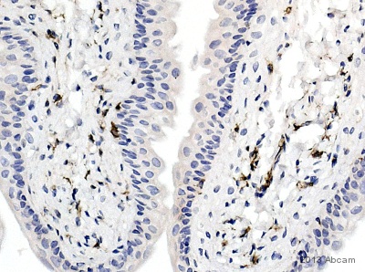 IHC-P image of Iba1 staining on marmoset bladder sections using ab5076 (1:1000). The sections were deparaffinized and subjected to heat mediated antigen retrieval using citric acid. The sections were blocked using 1% BSA for 10 mins at 21°C. ab5076 was diluted 1:1000 and incubted with the sections for 2 hours at 21°C. The secondary antibody used was Rabbit polyclonal to anti goat IgG conjugated to biotin (1:200)See Abreview
 ab5076 at 1/500 staining rat brain tissue sections by IHC-Fr. The tissue was paraformaldehyde fixed and a heat mediated antigen retrieval step was used, prior to incubation with the tissue for 30 minutes. An Alexa-Fluor ® 594 conjugated rabbit polyclonal antibody was used as the secondary.See Abreview
 ab5076 staining Iba1 in guinea pig brain tissue section by Immunohistochemistry (Formalin/PFA-fixed paraffin-embedded sections). Tissue underwent formaldehyde fixation before heat mediated antigen retrieval in sodium citrate and then blocking with 3% serum was performed for 30 minutes at RT. The primary antibody was used at dilution at 1/200 and incubated with sample at 2% blocking serum for 18 hours at 4°C. A Biotin conjugated horse polyclonal to goat IgG was used undiluted as secondary antibody.See Abreview
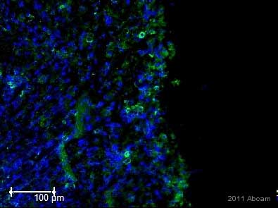
 AIF1 expression in an inflammatory foci in a skin biopsy. Paraffin embedded sections were deparaffinized and boiled in 10mM citrate buffer for 30 min in a microwave to expose the AIF1 antigen, and then blocked with 5% donkey serum for 20 min. Slides were incubated for 40 min with goat-anti-AIF1 antibody (1:100) and rinsed in three changes of PBS for 1 min each. A secondary antibody (donkey-anti-goat-FITC conjugated IgG; 1:50) was then applied to the slide for 40 min. Sections were washed again to remove the unbound antibody. The slides were counterstained with DaPI and viewed with a Nikon epi-fluorescent microscope. AIF1 positive cells appear green and the nuclei are stained blue. Numerous cells expressing the AIF1 protein are located around a vessel. (400X). Picture from Rreview by Carol Artlett submitted 30 July 2004.
 ab5076 at a 1/400 dilution staining rat brain tissue sections by Immunohistochemistry (frozen sections). The brain were frozen in isopentane, and the section postfixed with PFA2%. Following a short postfixation (15 minutes) the signal was very poor, while it improved after a longer fixation (1 hour). Heat mediated antigen retrieval was also attempted, but without a good result (excessive tissue specific aspecific binding, e.g. neurons), both on PFA postfixed frozen tissue and on tissue from perfused animals. An Alexa Fluor ® 568 conjugated antibody was used as the secondary antibody.This image is courtesy of an Abreview.See Abreview
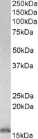 Anti-Iba1 antibody (ab5076) at 0.5 µg/ml + Rat Brain lysate in RIPA buffer at 35 µgdeveloped using the ECL technique
 ab5076 staining Iba1 in rat spleen tissue by Immunohistochemistry (Frozen sections).Tissue was fixed with formaldehyde, blocked using 10% serum for 30 minutes at 24°C, then incubated with ab5076 at a 1/500 dilution for 16 hours at 4°C. The secondary used was an Alexa Fluor 488 conjugated rabbit anti-goat polyclonal used at a 1/1000 dilution.See Abreview
 ab5076 staining Iba1 in rat glioblastoma cell line C6 by Immunocytochemistry/ Immunofluorescence.Cells were fixed in paraformaldehyde, permeabilized using 0,1% Triton X 100 in PBS, blocked with 0.5% BSA for 30 minutes at room temperature and then incubated with ab5076 at a 1/50 dilution for 16 hours at 4°C. The secondary used was a Cy3 conjugated rabbit anti-goat polyclonal used at a 1/120 dilution. Nuclei are counterstained with DAPI.See Abreview
Product References
Global changes in DNA methylation and hydroxymethylation in Alzheimer's disease - Global changes in DNA methylation and hydroxymethylation in Alzheimer's disease
Coppieters N, Dieriks BV, Lill C, Faull RL, Curtis MA, Dragunow M. Neurobiol Aging. 2014 Jun;35(6):1334-44. doi:
Glial activation in the early stages of brain metastasis: TSPO as a diagnostic - Glial activation in the early stages of brain metastasis: TSPO as a diagnostic
O'Brien ER, Kersemans V, Tredwell M, Checa B, Serres S, Soto MS, Gouverneur V, Leppert D, Anthony DC, Sibson NR. J Nucl Med. 2014 Feb;55(2):275-80.
T(2)-weighted MRI detects presymptomatic pathology in the SOD1 mouse model of - T(2)-weighted MRI detects presymptomatic pathology in the SOD1 mouse model of
Evans MC, Serres S, Khrapitchev AA, Stolp HB, Anthony DC, Talbot K, Turner MR, Sibson NR. J Cereb Blood Flow Metab. 2014 May;34(5):785-93.
PI3Kdelta inhibition reduces TNF secretion and neuroinflammation in a mouse - PI3Kdelta inhibition reduces TNF secretion and neuroinflammation in a mouse
Low PC, Manzanero S, Mohannak N, Narayana VK, Nguyen TH, Kvaskoff D, Brennan FH, Ruitenberg MJ, Gelderblom M, Magnus T, Kim HA, Broughton BR, Sobey CG, Vanhaesebroeck B, Stow JL, Arumugam TV, Meunier FA. Nat Commun. 2014 Mar 14;5:3450.
CXCL12 in astrocytes contributes to bone cancer pain through CXCR4-mediated - CXCL12 in astrocytes contributes to bone cancer pain through CXCR4-mediated
Shen W, Hu XM, Liu YN, Han Y, Chen LP, Wang CC, Song C. J Neuroinflammation. 2014 Apr 16;11:75.
Epidermal growth factor treatment of the adult brain subventricular zone leads to - Epidermal growth factor treatment of the adult brain subventricular zone leads to
Lindberg OR, Brederlau A, Kuhn HG. Stem Cell Reports. 2014 Mar 27;2(4):440-8.
Hyperlipidemic diet causes loss of olfactory sensory neurons, reduces olfactory - Hyperlipidemic diet causes loss of olfactory sensory neurons, reduces olfactory
Thiebaud N, Johnson MC, Butler JL, Bell GA, Ferguson KL, Fadool AR, Fadool JC, Gale AM, Gale DS, Fadool DA. J Neurosci. 2014 May 14;34(20):6970-84.
Transcription factors NRF2 and NF-kappaB are coordinated effectors of the Rho - Transcription factors NRF2 and NF-kappaB are coordinated effectors of the Rho
Cuadrado A, Martin-Moldes Z, Ye J, Lastres-Becker I. J Biol Chem. 2014 May 30;289(22):15244-58.
AAV-dominant negative tumor necrosis factor (DN-TNF) gene transfer to the - AAV-dominant negative tumor necrosis factor (DN-TNF) gene transfer to the
Alto LT, Chen X, Ruhn KA, Trevino I, Tansey MG. PLoS One. 2014 May 13;9(5):e96544.
Congenitally acquired persistent lymphocytic choriomeningitis viral infection - Congenitally acquired persistent lymphocytic choriomeningitis viral infection
Sun T, Vasek MJ, Klein RS. PLoS One. 2014 May 6;9(5):e96442.