Anti-Iba1 antibody
| Name | Anti-Iba1 antibody |
|---|---|
| Supplier | Abcam |
| Catalog | ab107159 |
| Prices | $392.00 |
| Sizes | 100 µg |
| Host | Goat |
| Clonality | Polyclonal |
| Isotype | IgG |
| Applications | IHC-F IHC-P WB |
| Species Reactivities | Mouse, Rat, Dog, Human, Marmoset, Pig, Monkey, Hamster |
| Antigen | Synthetic peptide conjugated to KLH derived from within residues 100 to the C-terminus of Rat Iba1 |
| Description | Goat Polyclonal |
| Gene | AIF1 |
| Conjugate | Unconjugated |
| Supplier Page | Shop |
Product images
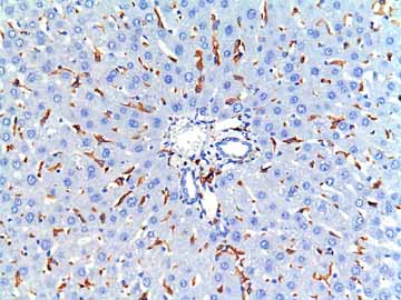 ab107159 staining Iba1 in Rat liver tissue sections by Immunohistochemistry (IHC-P - paraformaldehyde-fixed, paraffin-embedded sections). Tissue was fixed with formaldehyde and blocked with 1% BSA for 15 minutes at 20°C; antigen retrieval was by heat mediation in a citrate buffer. Samples were incubated with primary antibody (1/2000 in PBS + 1% BSA) for 1 hour at 20°C. An undiluted HRP-conjugated anti-goat IgG polyclonal was used as the secondary antibody. Kupffer cells are positive for Iba-1.See Abreview
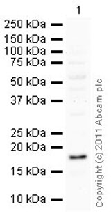 Anti-Iba1 antibody (ab107159) at 1 µg/ml + Rat Cortex Tissue Lysate at 10 µgSecondaryRabbit polyclonal to Goat IgG - H&L - Pre-Adsorbed (HRP) at 1/3000 dilutiondeveloped using the ECL techniquePerformed under reducing conditions.
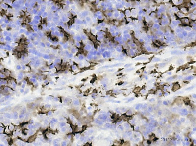
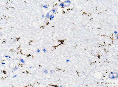 IHC-P image of Iba1 staining on human brain sections using ab107159 (1:2000). The paraffin embedded sectons were subjected to heat mediated antigen retrieval using citric acid. The sections was then blocked using 1% BSA at 21°C for 10 min. The secondary antibody was horse polyclonal to anti-sheep conjugated to biotin at 1:200.See Abreview
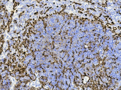 IHC-P image of Iba1 staining on mouse spleen sections using ab107159 (1:2000). The paraffin embedded sectons were subjected to heat mediated antigen retrieval using citric acid. The sections was then blocked using 1% BSA at 21°C for 10 min. The secondary antibody was horse polyclonal to anti-sheep conjugated to biotin at 1:200.See Abreview
 IHC-P image of Iba1 staining on rat spleen sections using ab107159 (1:2000). The paraffin embedded sectons were subjected to heat mediated antigen retrieval using citric acid. The sections was then blocked using 1% BSA at 21°C for 10 min. The primary antibody was incubated at 21°C for 2 hours. The secondary antibody was goat polyclonal to anti-sheep conjugated to biotin at 1:250.See Abreview
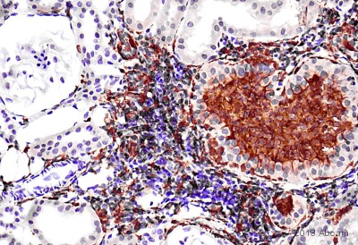 IHC-P image of Iba1 staining on marmoset (common) kidney sections using ab107159 (1:5000). The image shows macrophage infiltration in the cortex. The paraffin embedded sectons were subjected to heat mediated antigen retrieval using citric acid. The sections was then blocked using 1% BSA at 21°C for 10 min. The primary antibody was incubated at 21°C for 16 hours. The secondary antibody was rabbit polyclonal to sheep IgG conjugated to biotin at 1:200.See Abreview
Product References
Extra-prostatic transgene-associated neoplastic lesions in transgenic - Extra-prostatic transgene-associated neoplastic lesions in transgenic
Berman-Booty LD, Thomas-Ahner JM, Bolon B, Oglesbee MJ, Clinton SK, Kulp SK, Chen CS, La Perle KM. Toxicol Pathol. 2015 Feb;43(2):186-97.
Marmosets as a preclinical model for testing "off-label" use of doxycycline to - Marmosets as a preclinical model for testing "off-label" use of doxycycline to
VanderVeen N, Paran C, Appelhans A, Krasinkiewicz J, Lemons R, Appelman H, Doherty R, Palmer D, Ng P, Lowenstein PR, Castro MG. Mol Ther Methods Clin Dev. 2014 Feb 5;1. pii: 10.
New alpha- and gamma-synuclein immunopathological lesions in human brain. - New alpha- and gamma-synuclein immunopathological lesions in human brain.
Surgucheva I, Newell KL, Burns J, Surguchov A. Acta Neuropathol Commun. 2014 Sep 11;2:132.
Isoflurane on brain inflammation. - Isoflurane on brain inflammation.
Altay O, Suzuki H, Hasegawa Y, Ostrowski RP, Tang J, Zhang JH. Neurobiol Dis. 2014 Feb;62:365-71.
General anesthetics inhibit LPS-induced IL-1beta expression in glial cells. - General anesthetics inhibit LPS-induced IL-1beta expression in glial cells.
Tanaka T, Kai S, Matsuyama T, Adachi T, Fukuda K, Hirota K. PLoS One. 2013 Dec 11;8(12):e82930.