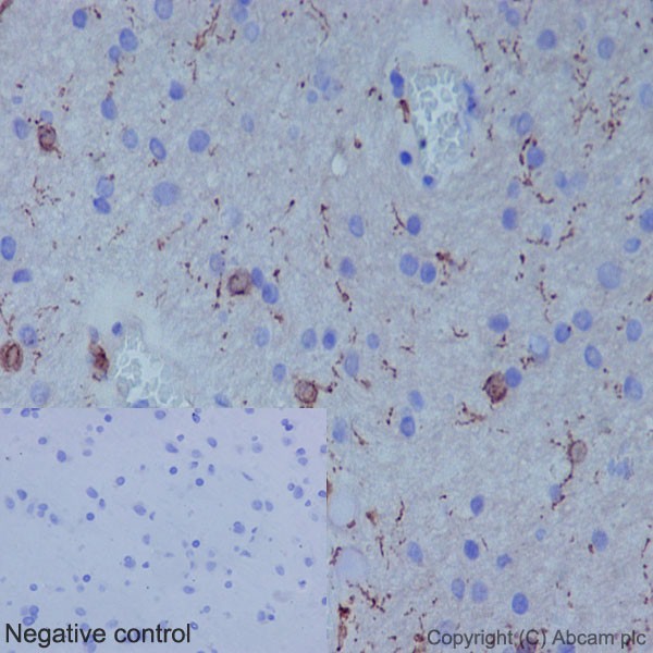
Immunohistochemical analysis of paraffin-embedded Human cerebral cortex tissue labeling Iba1 with ab178846 at a 1/2000 dilution showing cytoplasm and nuclear staining on Glial cells. Counter stained with hematoxylin. Prediluted HRP Polymer for Rabbit/Mouse IgG was used as the secondary aantibody. Negative control also shown.
![All lanes : Anti-Iba1 antibody [EPR16588] (ab178846) at 1/10000 dilutionLane 1 : THP-1 (Human monocytic leukemia cells) whole cell lysateLane 2 : U937 (Human histiocytic lymphoma cells) whole cell lysateLane 3 : Human spleen whole cell lysateLysates/proteins at 10 µg per lane.SecondaryGoat Anti-Rabbit IgG, (H+L), Peroxidase conjugated at 1/1000 dilutiondeveloped using the ECL technique](http://www.bioprodhub.com/system/product_images/ab_products/2/sub_3/6066_ab178846-228130-ab178846wb2.jpg)
All lanes : Anti-Iba1 antibody [EPR16588] (ab178846) at 1/10000 dilutionLane 1 : THP-1 (Human monocytic leukemia cells) whole cell lysateLane 2 : U937 (Human histiocytic lymphoma cells) whole cell lysateLane 3 : Human spleen whole cell lysateLysates/proteins at 10 µg per lane.SecondaryGoat Anti-Rabbit IgG, (H+L), Peroxidase conjugated at 1/1000 dilutiondeveloped using the ECL technique
![Anti-Iba1 antibody [EPR16588] (ab178846) at 1/2000 dilution + HL-60 (Human promyelocytic leukemia cells) whole cell lysate at 10 µgSecondaryGoat Anti-Rabbit IgG, (H+L), Peroxidase conjugated at 1/1000 dilution](http://www.bioprodhub.com/system/product_images/ab_products/2/sub_3/6067_ab178846-228124-ab178846wb1.jpg)
Anti-Iba1 antibody [EPR16588] (ab178846) at 1/2000 dilution + HL-60 (Human promyelocytic leukemia cells) whole cell lysate at 10 µgSecondaryGoat Anti-Rabbit IgG, (H+L), Peroxidase conjugated at 1/1000 dilution

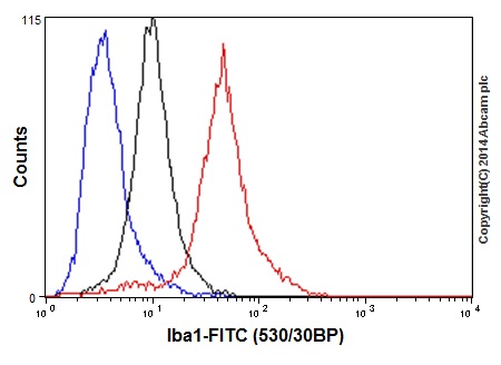
Flow cytometry analysis of 2% paraformaldehyde fixed U937 (Human histiocytic lymphoma cells) cells labeling Iba1 with ab178846 at 1/160 dilution (red line). Secondary antibody used is a goat anti rabbit IgG (FITC) at 1/150 dilution. The isotype control is rabbit monoclonal IgG (black line). The unlabeled control is cells without incubation with primary and secondary antibodies (blue line).

![All lanes : Anti-Iba1 antibody [EPR16588] (ab178846) at 1/10000 dilutionLane 1 : THP-1 (Human monocytic leukemia cells) whole cell lysateLane 2 : U937 (Human histiocytic lymphoma cells) whole cell lysateLane 3 : Human spleen whole cell lysateLysates/proteins at 10 µg per lane.SecondaryGoat Anti-Rabbit IgG, (H+L), Peroxidase conjugated at 1/1000 dilutiondeveloped using the ECL technique](http://www.bioprodhub.com/system/product_images/ab_products/2/sub_3/6066_ab178846-228130-ab178846wb2.jpg)
![Anti-Iba1 antibody [EPR16588] (ab178846) at 1/2000 dilution + HL-60 (Human promyelocytic leukemia cells) whole cell lysate at 10 µgSecondaryGoat Anti-Rabbit IgG, (H+L), Peroxidase conjugated at 1/1000 dilution](http://www.bioprodhub.com/system/product_images/ab_products/2/sub_3/6067_ab178846-228124-ab178846wb1.jpg)
