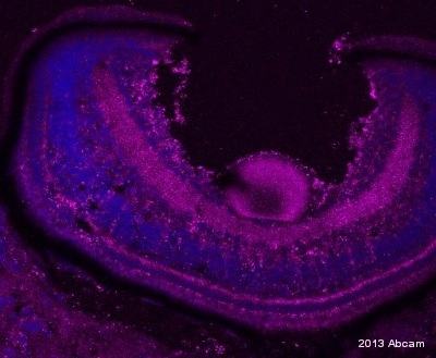
ab118032 staining ID3 in zebrafish retina sections by IHC-Fr. The tissue was fixed with paraformaldehyde and an antigen retrieval step was performed with sodium citrate pH 6. Blocking of the sample was done with 5% BSA in PBS containing 01% Tween 20 and 0.5% Triton X, for 60 minutes at 23°C, followed by staining with ab118032 at 1/100 in blocking solution for 16h at 4°C. An alexa 647 conjugated goat anti-mouset polyclonal antibody at 1/1000 was used as the secondary antibody. Nuclei are stained in blue with DAPI. ID3 expression can be observed in Muller cells.See Abreview
![All lanes : Anti-ID3 antibody [10D3] (ab118032) at 1/500 dilutionLane 1 : HEK293T cell lysate transfected with pCMV6-ENTRY control cDNALane 2 : HEK293T cell lysate transfected with pCMV6-ENTRY ID3 cDNA Lysates/proteins at 5 µg per lane.](http://www.bioprodhub.com/system/product_images/ab_products/2/sub_3/6312_ID3-Primary-antibodies-ab118032-1.jpg)
All lanes : Anti-ID3 antibody [10D3] (ab118032) at 1/500 dilutionLane 1 : HEK293T cell lysate transfected with pCMV6-ENTRY control cDNALane 2 : HEK293T cell lysate transfected with pCMV6-ENTRY ID3 cDNA Lysates/proteins at 5 µg per lane.

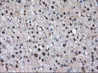
ab118032 at 1/50 dilution staining ID3 in Paraffin-embedded Human Liver tissue by Immunohistochemistry.

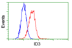
ab118032 at 1/100 dilution staining ID3 in HeLa cells by Flow cytometry (Red) compared to a nonspecific negative control antibody (Blue).

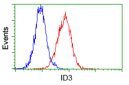
ab118032 at 1/100 dilution staining ID3 in Jurkat cells by Flow cytometry (Red) compared to a nonspecific negative control antibody (Blue).

![All lanes : Anti-ID3 antibody [10D3] (ab118032) at 1/500 dilutionLane 1 : HEK293T cell lysate transfected with pCMV6-ENTRY control cDNALane 2 : HEK293T cell lysate transfected with pCMV6-ENTRY ID3 cDNA Lysates/proteins at 5 µg per lane.](http://www.bioprodhub.com/system/product_images/ab_products/2/sub_3/6312_ID3-Primary-antibodies-ab118032-1.jpg)


