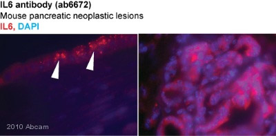
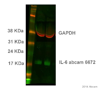
Anti-IL6 antibody
| Name | Anti-IL6 antibody |
|---|---|
| Supplier | Abcam |
| Catalog | ab6672 |
| Host | Rabbit |
| Clonality | Polyclonal |
| Isotype | IgG |
| Applications | WB ELISA IHC-P RIA IP IHC-F B/N ICC/IF ICC/IF |
| Species Reactivities | Mouse, Rat, Human, Pig |
| Antigen | recombinant human IL-6 produced in E |
| Description | Rabbit Polyclonal |
| Gene | IL6 |
| Conjugate | Unconjugated |
| Supplier Page | Shop |
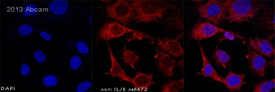
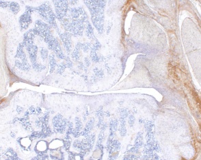
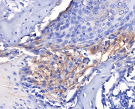
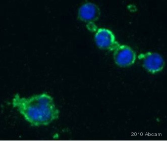
Product images
Product References
Pyrrolidine dithiocarbamate attenuates surgery-induced neuroinflammation and - Pyrrolidine dithiocarbamate attenuates surgery-induced neuroinflammation and
Zhang J, Jiang W, Zuo Z. Neuroscience. 2014 Mar 7;261:1-10.
Pro-inflammatory mediators and apoptosis correlate to rt-PA response in a novel - Pro-inflammatory mediators and apoptosis correlate to rt-PA response in a novel
Ansar S, Chatzikonstantinou E, Thiagarajah R, Tritschler L, Fatar M, Hennerici MG, Meairs S. PLoS One. 2014 Jan 20;9(1):e85849.
Clearance of senescent hepatocytes in a neoplastic-prone microenvironment delays - Clearance of senescent hepatocytes in a neoplastic-prone microenvironment delays
Marongiu F, Serra MP, Sini M, Angius F, Laconi E. Aging (Albany NY). 2014 Jan;6(1):26-34.
IL-6 regulates extracellular matrix remodeling associated with aortic dilation in - IL-6 regulates extracellular matrix remodeling associated with aortic dilation in
Ju X, Ijaz T, Sun H, Lejeune W, Vargas G, Shilagard T, Recinos A 3rd, Milewicz DM, Brasier AR, Tilton RG. J Am Heart Assoc. 2014 Jan 21;3(1):e000476.
Role of unphosphorylated transcription factor STAT3 in late cerebral ischemia - Role of unphosphorylated transcription factor STAT3 in late cerebral ischemia
Samraj AK, Muller AH, Grell AS, Edvinsson L. J Cereb Blood Flow Metab. 2014 May;34(5):759-63.
AIM2 mediates inflammation-associated renal damage in hepatitis B - AIM2 mediates inflammation-associated renal damage in hepatitis B
Zhen J, Zhang L, Pan J, Ma S, Yu X, Li X, Chen S, Du W. Mediators Inflamm. 2014;2014:190860.
Sophocarpine attenuates liver fibrosis by inhibiting the TLR4 signaling pathway - Sophocarpine attenuates liver fibrosis by inhibiting the TLR4 signaling pathway
Qian H, Shi J, Fan TT, Lv J, Chen SW, Song CY, Zheng ZW, Xie WF, Chen YX. World J Gastroenterol. 2014 Feb 21;20(7):1822-32.
Perineural dexmedetomidine attenuates inflammation in rat sciatic nerve via the - Perineural dexmedetomidine attenuates inflammation in rat sciatic nerve via the
Huang Y, Lu Y, Zhang L, Yan J, Jiang J, Jiang H. Int J Mol Sci. 2014 Mar 6;15(3):4049-59.
Interleukin-6 secretion by astrocytes is dynamically regulated by - Interleukin-6 secretion by astrocytes is dynamically regulated by
Codeluppi S, Fernandez-Zafra T, Sandor K, Kjell J, Liu Q, Abrams M, Olson L, Gray NS, Svensson CI, Uhlen P. PLoS One. 2014 Mar 25;9(3):e92649.
Synthesis of IL-6 by hepatocytes is a normal response to common hepatic stimuli. - Synthesis of IL-6 by hepatocytes is a normal response to common hepatic stimuli.
Norris CA, He M, Kang LI, Ding MQ, Radder JE, Haynes MM, Yang Y, Paranjpe S, Bowen WC, Orr A, Michalopoulos GK, Stolz DB, Mars WM. PLoS One. 2014 Apr 24;9(4):e96053.
