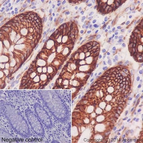
Immunohistochemical analysis of paraffin-embedded Human colon tissue labeling Integrin alpha 2 with ab181548 at 1/500 dilution followed by Goat Anti-Rabbit HRP (IgG H&L) (ab97051) at 1/500 dilution. Membrane and weak cytoplasmic staining on epithelial cells of human colon is observed. Counter stained with Hematoxylin.Negative control: Used PBS instead of primary antibody.
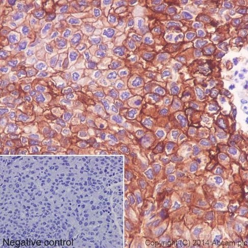
Immunohistochemical analysis of paraffin-embedded Human squamous cell carcinoma of cervix tissue labeling Integrin alpha 2 with ab181548 at 1/500 dilution followed by Goat Anti-Rabbit HRP (IgG H&L) (ab97051) at 1/500 dilution. Membrane and weak cytoplasmic staining on epithelial cells of human squamous cell carcinoma of cervix tissue is observed. Counter stained with Hematoxylin.Negative control: Used PBS instead of primary antibody.
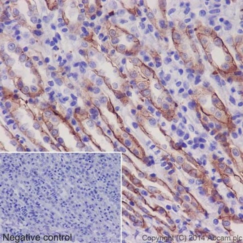
Immunohistochemical analysis of paraffin-embedded Mouse kidney tissue labeling Integrin alpha 2 with ab181548 at 1/500 dilution followed by Goat Anti-Rabbit HRP (IgG H&L) (ab97051) at 1/500 dilution. Membrane and weak cytoplasmic staining on epithelial cells of Mouse kidney tubule is observed. Counter stained with Hematoxylin.Negative control: Used PBS instead of primary antibody.
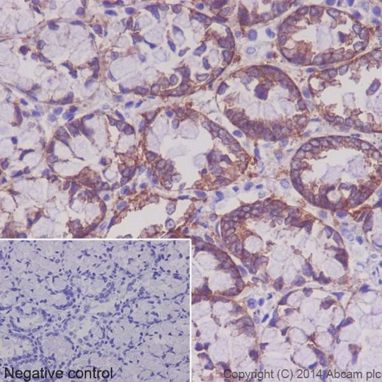
Immunohistochemical analysis of paraffin-embedded Rat colon tissue labeling Integrin alpha 2 with ab181548 at 1/500 dilution followed by Goat Anti-Rabbit HRP (IgG H&L) (ab97051) at 1/500 dilution. Membrane staining on epithelial cells of Rat colon tissue is observed. Counter stained with Hematoxylin.Negative control: Used PBS instead of primary antibody.
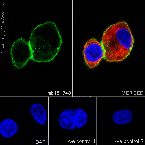
Immunofluorescent analysis of 4% paraformaldehyde-fixed, 0.1% Triton X-100 permeabilized PC-3 (Human prostate adenocarcinoma cell line) cells labeling integrin alpha 2 with ab181548 at 1/100 dilution, followed by Goat anti-rabbit IAlexa Fluor® 488 (IgG) (ab150077) secondary antibody at 1/400 dilution (green). Confocal image showing membrane and weakly cytoplasmic staining on PC-3 cell line is observed. The nuclear counterstain is DAPI (blue). Tubulin is detected with ab7291 (anti-Tubulin mouse mAb) at 1/500 dilution and ab150120 (goat anti-mouse AlexaFluor®594 secondary antibody) at 1/500 dilution (red).The negative controls are as follows:--ve control 1 - ab181548 at 1/100 dilution followed by ab150120 (AlexaFluor®594 Goat anti-Mouse secondary) at 1/500 dilution.-ve control 2. - ab7291 (anti-Tubulin mouse mAb) at 1/500 dilution followed by ab150077 (Alexa Fluor®488 Goat Anti-Rabbit IgG H&L) at 1/400 dilution.
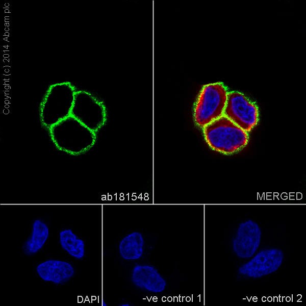
Immunofluorescent analysis of 4% paraformaldehyde-fixed, 0.1% Triton X-100 permeabilized MCF7 (Human breast adenocarcinoma cell line) cells labeling integrin alpha 2 with ab181548 at 1/100 dilution, followed by Goat anti-rabbit IAlexa Fluor® 488 (IgG) (ab150077) secondary antibody at 1/400 dilution (green). Confocal image showing membrane staining on MCF7 cell line is observed. The nuclear counterstain is DAPI (blue). Tubulin is detected with ab7291 (anti-Tubulin mouse mAb) at 1/500 dilution and ab150120 (goat anti-mouse AlexaFluor®594 secondary antibody) at 1/500 dilution (red).The negative controls are as follows:--ve control 1 - ab181548 at 1/100 dilution followed by ab150120 (AlexaFluor®594 Goat anti-Mouse secondary) at 1/500 dilution.-ve control 2. - ab7291 (anti-Tubulin mouse mAb) at 1/500 dilution followed by ab150077 (Alexa Fluor®488 Goat Anti-Rabbit IgG H&L) at 1/400 dilution.
![All lanes : Anti-Integrin alpha 2 antibody [EPR17338] - C-terminal (ab181548) at 1/20000 dilutionLane 1 : A549 (Human lung carcinoma) whole cell lysatesLane 2 : A431 (Human epidermoid carcinoma) whole cell lysatesLane 3 : 293T (Human epithelial cells from embryonic kidney) whole cell lysatesLane 4 : T-47D (Human ductal breast epithelial tumor cell line) whole cell lysatesLysates/proteins at 20 µg per lane.SecondaryGoat Anti-Rabbit IgG, (H+L),Peroxidase conjugated at 1/1000 dilution](http://www.bioprodhub.com/system/product_images/ab_products/2/sub_3/8948_ab181548-234862-IntergrinWB1.jpg)
All lanes : Anti-Integrin alpha 2 antibody [EPR17338] - C-terminal (ab181548) at 1/20000 dilutionLane 1 : A549 (Human lung carcinoma) whole cell lysatesLane 2 : A431 (Human epidermoid carcinoma) whole cell lysatesLane 3 : 293T (Human epithelial cells from embryonic kidney) whole cell lysatesLane 4 : T-47D (Human ductal breast epithelial tumor cell line) whole cell lysatesLysates/proteins at 20 µg per lane.SecondaryGoat Anti-Rabbit IgG, (H+L),Peroxidase conjugated at 1/1000 dilution
![All lanes : Anti-Integrin alpha 2 antibody [EPR17338] - C-terminal (ab181548) at 1/5000 dilutionLane 1 : Human fetal brain whole cell lysatesLane 2 : Human fetal heart whole cell lysatesLysates/proteins at 10 µg per lane.SecondaryAnti-Rabbit IgG (HRP), specific to the non-reduced form of IgG at 1/1000 dilution](http://www.bioprodhub.com/system/product_images/ab_products/2/sub_3/8949_ab181548-234863-integrinWB2.jpg)
All lanes : Anti-Integrin alpha 2 antibody [EPR17338] - C-terminal (ab181548) at 1/5000 dilutionLane 1 : Human fetal brain whole cell lysatesLane 2 : Human fetal heart whole cell lysatesLysates/proteins at 10 µg per lane.SecondaryAnti-Rabbit IgG (HRP), specific to the non-reduced form of IgG at 1/1000 dilution
![All lanes : Anti-Integrin alpha 2 antibody [EPR17338] - C-terminal (ab181548) at 1/5000 dilutionLane 1 : Mouse heart tissue lysateLane 2 : Mouse kidney tissue lysateLane 3 : Rat spleen tissue lysateLane 4 : C6 (Rat glial tumor cells) whole cell lysateLane 5 : NIH/3T3 (Mouse embyro fibroblast cells) whole cell lysateLysates/proteins at 10 µg per lane.SecondaryGoat Anti-Rabbit IgG, (H+L),Peroxidase conjugated at 1/1000 dilution](http://www.bioprodhub.com/system/product_images/ab_products/2/sub_3/8950_ab181548-234872-integrinWB3.jpg)
All lanes : Anti-Integrin alpha 2 antibody [EPR17338] - C-terminal (ab181548) at 1/5000 dilutionLane 1 : Mouse heart tissue lysateLane 2 : Mouse kidney tissue lysateLane 3 : Rat spleen tissue lysateLane 4 : C6 (Rat glial tumor cells) whole cell lysateLane 5 : NIH/3T3 (Mouse embyro fibroblast cells) whole cell lysateLysates/proteins at 10 µg per lane.SecondaryGoat Anti-Rabbit IgG, (H+L),Peroxidase conjugated at 1/1000 dilution
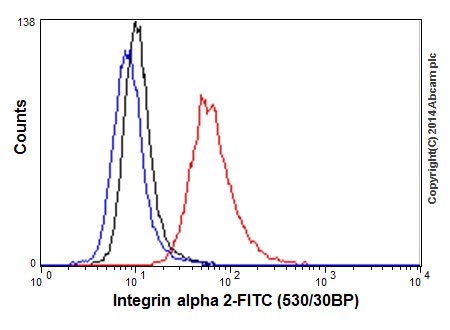
Flow cytometric analysis of 2% paraformaldehyde-fixed A549 (Human lung carcinoma) cells labeling integrin alpha 2 with ab181549 at 1/160 dilution (red) compared with a rabbit monoclonal IgG isotype control (black) and a unlabelled control (cells without incubation with primary antibody and secondary antibody; blue). Goat anti rabbit IgG (FITC) at 1/150 dilution was used as the secondary antibody.
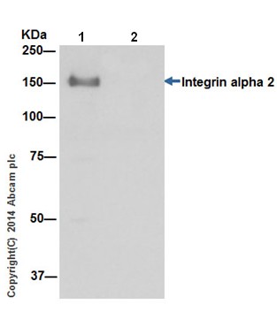
Integrin alpha 2 was immunoprecipitated from 1mg of T-47D (Human ductal breast epithelial tumor cell line) whole cell extract with ab181548 at 1/150 dilution. Western blot was performed using ab181548 at 1/20,000 dilution. Anti-Rabbit IgG (HRP), specific to the non-reduced form of IgG, was used as secondary antibody at 1/1500 dilution. Lane 1: T-47D whole cell extract Lane 2: PBS instead of T-47D whole cell extract.Blocking and dilution buffer and concentration: 5% NFDM/TBST.