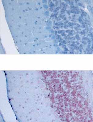
ab52012 staining KALRN in adult brain tissue by Immunohistochemistry (paraffin embedded sections). Tissue was fixed with paraformaldehyde and a heat mediated antigen retrieval step performed using citrate buffer. Tissues were then blocked with 15% serum for 45 minutes at 20°C followed by incubation with the primary antibody, at a 1/250 dilution, for 24 hours at 4°C. A Biotin-conjugated rabbit anti-goat IgG was used as secondary antibody at a 1/250 dilution.Upper image: Un-treated.Lower image: Treated with peptide to KALRN.Counterstained with Hämalaun.See Abreview

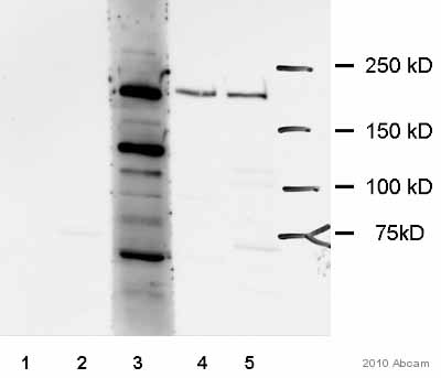
All lanes : Anti-KALRN antibody (ab52012) at 1/500 dilutionLane 1 : Whole tissue lysate prepared from mouse brain, treated with peptide.Lane 2 : Whole cell lysate prepared from mouse brain cells over-expressing KALRN (with FLAG-tag) treated with peptide.Lane 3 : Whole cell lysate prepared from mouse brain cells over-expressing KALRN, with FLAG-tag, detected with anti-FLAG antibody.Lane 4 : Whole tissue lysate prepared from mouse brainLane 5 : Whole cell lysate prepared from mouse brain cells over-expressing KALRN, with FLAG-tag.Lysates/proteins at 30 µg per lane.SecondaryDonkey anti-goat IgG conjugated to HRP at 1/10000 dilutiondeveloped using the ECL techniquePerformed under non-reducing conditions.

Anti-KALRN antibody (ab52012) at 0.5 µg/ml + Mouse Brain lysate in RIPA buffer at 35 µgdeveloped using the ECL techniquePerformed under reducing conditions.


