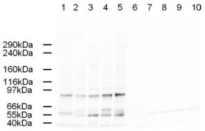
Western blot using Affinity Purified anti-AP2A antibody shows detection of a band just below 100 kDa corresponding to Human AP2A1 in a various preparations. Lane 1 - HeLa nuclear extract, Lane 2 - HeLa, Lane 3 - 293, Lane 4 - A431 and Lane 5 - Jurkat whole cell lysates. In lanes 6-10 the antibody was preincubated with 1 µg/ml of the immunizing peptide which effectively blocks the specific reactivity of this antibody with AP2A. Approximately 20 µg of each lysate was run on a SDS-PAGE and transferred onto nitrocellulose followed by reaction with a 1:500 dilution of anti-AP2A antibody. Detection occurred using a 1:5,000 dilution of HRP-labeled Rabbit anti-Goat IgG for 1 hour at room temperature. A chemi-luminescence system was used for signal detection (Roche) using a 60-sec exposure time.