Anti-KAT13A / SRC1 antibody [1135/H4] - ChIP Grade
| Name | Anti-KAT13A / SRC1 antibody [1135/H4] - ChIP Grade |
|---|---|
| Supplier | Abcam |
| Catalog | ab84 |
| Prices | $400.00 |
| Sizes | 50 µg |
| Host | Mouse |
| Clonality | Monoclonal |
| Isotype | IgG1 |
| Clone | 1135/H4 |
| Applications | IHC-P ChIP FC WB IP |
| Species Reactivities | Mouse, Rat, Human, Monkey |
| Antigen | Fusion protein, corresponding to amino acids 477-947 of Human SRC1 |
| Description | Mouse Monoclonal |
| Gene | NCOA1 |
| Conjugate | Unconjugated |
| Supplier Page | Shop |
Product images
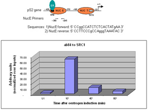 Sonicated Chromatin prepared from untreated (UI) or 17beta-estradiol (E2) treated MCF7 cells was subjected to the ChIP procedure with ab84 to SRC1 and the immunoprecipitated chromatin was analysed in the proximal region of the estrogen-responsive pS2 promoter (as shown above) and quantified by real-time PCR (values are nomalized over inputs). The primers are designed to follow the nucleosome E (including the Estrogen Responsive Element ERE). 5 µl of ab84 and 2x10⁶ cells were used in each ChIP experiment.
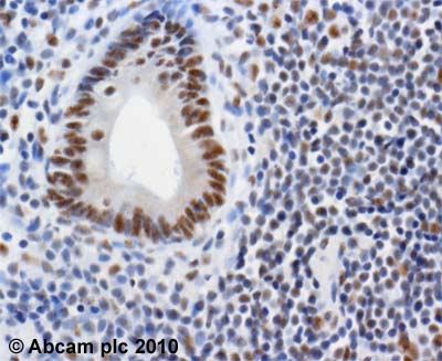 ab84 (2µg/ml) staining SRC1 in human colonic mucosa using an automated system (DAKO Autostainer Plus). Using this protocol there is strong nuclear staining of mucosal epithelium and lymphocytes.Sections were rehydrated and antigen retrieved with the Dako 3 in 1 AR buffer EDTA pH 9.0 in a DAKO PT link. Slides were peroxidase blocked in 3% H2O2 in methanol for 10 mins. They were then blocked with Dako Protein block for 10 minutes (containing casein 0.25% in PBS) then incubated with primary antibody for 20 min and detected with Dako envision flex amplification kit for 30 minutes. Colorimetric detection was completed with Diaminobenzidine for 5 minutes. Slides were counterstained with Haematoxylin and coverslipped under DePeX. Please note that, for manual staining, optimization of primary antibody concentration and incubation time is recommended. Signal amplification may be required.
![Overlay histogram showing HeLa cells stained with ab84 (red line). The cells were fixed with 80% methanol (5 min) and then permeabilized with 0.1% PBS-Tween for 20 min. The cells were then incubated in 1x PBS / 10% normal goat serum / 0.3M glycine to block non-specific protein-protein interactions. The cells were then incubated with the antibody (ab84, 1µg/1x106 cells) for 30 min at 22ºC. The secondary antibody used was DyLight® 488 goat anti-mouse IgG (H+L) (ab96879) at 1/500 dilution for 30 min at 22ºC. Isotype control antibody (black line) was mouse IgG1 [ICIGG1] (ab91353, 2µg/1x106 cells ) used under the same conditions. Acquisition of >5,000 events was performed. This antibody gave a positive signal in HeLa cells fixed with 4% paraformaldehyde (10 min)/permeabilized in 0.1% PBS-Tween used under the same conditions.](http://www.bioprodhub.com/system/product_images/ab_products/2/sub_3/11442_KAT13A-SRC1-Primary-antibodies-ab84-2.jpg) Overlay histogram showing HeLa cells stained with ab84 (red line). The cells were fixed with 80% methanol (5 min) and then permeabilized with 0.1% PBS-Tween for 20 min. The cells were then incubated in 1x PBS / 10% normal goat serum / 0.3M glycine to block non-specific protein-protein interactions. The cells were then incubated with the antibody (ab84, 1µg/1x106 cells) for 30 min at 22ºC. The secondary antibody used was DyLight® 488 goat anti-mouse IgG (H+L) (ab96879) at 1/500 dilution for 30 min at 22ºC. Isotype control antibody (black line) was mouse IgG1 [ICIGG1] (ab91353, 2µg/1x106 cells ) used under the same conditions. Acquisition of >5,000 events was performed. This antibody gave a positive signal in HeLa cells fixed with 4% paraformaldehyde (10 min)/permeabilized in 0.1% PBS-Tween used under the same conditions.
Overlay histogram showing HeLa cells stained with ab84 (red line). The cells were fixed with 80% methanol (5 min) and then permeabilized with 0.1% PBS-Tween for 20 min. The cells were then incubated in 1x PBS / 10% normal goat serum / 0.3M glycine to block non-specific protein-protein interactions. The cells were then incubated with the antibody (ab84, 1µg/1x106 cells) for 30 min at 22ºC. The secondary antibody used was DyLight® 488 goat anti-mouse IgG (H+L) (ab96879) at 1/500 dilution for 30 min at 22ºC. Isotype control antibody (black line) was mouse IgG1 [ICIGG1] (ab91353, 2µg/1x106 cells ) used under the same conditions. Acquisition of >5,000 events was performed. This antibody gave a positive signal in HeLa cells fixed with 4% paraformaldehyde (10 min)/permeabilized in 0.1% PBS-Tween used under the same conditions.
Product References
Cyclic AMP enhances progesterone action in human myometrial cells. - Cyclic AMP enhances progesterone action in human myometrial cells.
Chen L, Lei K, Malawana J, Yulia A, Sooranna SR, Bennett PR, Liang Z, Grammatopoulos D, Johnson MR. Mol Cell Endocrinol. 2014 Jan 25;382(1):334-43.
Genome-wide analysis of binding sites and direct target genes of the orphan - Genome-wide analysis of binding sites and direct target genes of the orphan
Montemayor C, Montemayor OA, Ridgeway A, Lin F, Wheeler DA, Pletcher SD, Pereira FA. PLoS One. 2010 Jan 27;5(1):e8910.
Papillary and muscle invasive bladder tumors with distinct genomic stability - Papillary and muscle invasive bladder tumors with distinct genomic stability
Bentley J, L'Hote C, Platt F, Hurst CD, Lowery J, Taylor C, Sak SC, Harnden P, Knowles MA, Kiltie AE. Genes Chromosomes Cancer. 2009 Apr;48(4):310-21.
Progesterone induction of the 11beta-hydroxysteroid dehydrogenase type 2 promoter - Progesterone induction of the 11beta-hydroxysteroid dehydrogenase type 2 promoter
Subtil-Rodriguez A, Millan-Arino L, Quiles I, Ballare C, Beato M, Jordan A. Mol Cell Biol. 2008 Jun;28(11):3830-49.
4-Hydroxy-PCB106 acts as a direct thyroid hormone receptor agonist in rat GH3 - 4-Hydroxy-PCB106 acts as a direct thyroid hormone receptor agonist in rat GH3
You SH, Gauger KJ, Bansal R, Zoeller RT. Mol Cell Endocrinol. 2006 Sep 26;257-258:26-34. Epub 2006 Aug 23.