Anti-LAP1 antibody [RL13]
| Name | Anti-LAP1 antibody [RL13] |
|---|---|
| Supplier | Abcam |
| Catalog | ab2737 |
| Prices | $370.00 |
| Sizes | 200 µl |
| Host | Mouse |
| Clonality | Monoclonal |
| Isotype | IgG1 |
| Clone | RL13 |
| Applications | IHC-P ICC/IF ICC/IF IP WB |
| Species Reactivities | Mouse, Rat, Human |
| Antigen | Other Immunogen Type corresponding to Rat LAP1 |
| Description | Mouse Monoclonal |
| Gene | LRRC7 |
| Conjugate | Unconjugated |
| Supplier Page | Shop |
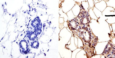
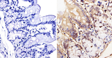
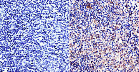
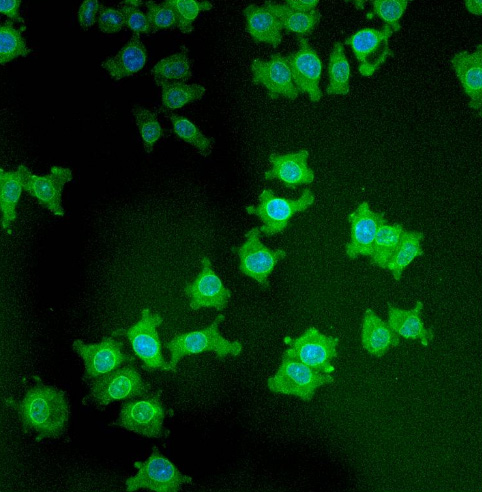
Product images
Product References
Distinctive T cell-suppressive signals from nuclearized type 1 sphingosine - Distinctive T cell-suppressive signals from nuclearized type 1 sphingosine
Liao JJ, Huang MC, Graler M, Huang Y, Qiu H, Goetzl EJ. J Biol Chem. 2007 Jan 19;282(3):1964-72. Epub 2006 Nov 22.
Lamin-binding fragment of LAP2 inhibits increase in nuclear volume during the - Lamin-binding fragment of LAP2 inhibits increase in nuclear volume during the
Yang L, Guan T, Gerace L. J Cell Biol. 1997 Dec 1;139(5):1077-87.
Structural analysis of the p62 complex, an assembly of O-linked glycoproteins - Structural analysis of the p62 complex, an assembly of O-linked glycoproteins
Guan T, Muller S, Klier G, Pante N, Blevitt JM, Haner M, Paschal B, Aebi U, Gerace L. Mol Biol Cell. 1995 Nov;6(11):1591-603.
cDNA cloning and characterization of lamina-associated polypeptide 1C (LAP1C), an - cDNA cloning and characterization of lamina-associated polypeptide 1C (LAP1C), an
Martin L, Crimaudo C, Gerace L. J Biol Chem. 1995 Apr 14;270(15):8822-8.
Integral membrane proteins of the nuclear envelope interact with lamins and - Integral membrane proteins of the nuclear envelope interact with lamins and
Foisner R, Gerace L. Cell. 1993 Jul 2;73(7):1267-79.
Integral membrane proteins specific to the inner nuclear membrane and associated - Integral membrane proteins specific to the inner nuclear membrane and associated
Senior A, Gerace L. J Cell Biol. 1988 Dec;107(6 Pt 1):2029-36.


