![All lanes : Anti-LOX antibody [EPR4025] (ab174316) at 1/3700 dilution (purified)Lane 1 : WI-38 cell lysateLane 2 : Jurkat cell lysateLane 3 : Mouse brainLane 4 : Rat brainLysates/proteins at 20 µg per lane.SecondaryHRP goat anti-rabbit (H+L) at 1/1000 dilution](http://www.bioprodhub.com/system/product_images/ab_products/2/sub_3/17038_ab174316-239764-174316-WB-2.jpg)
All lanes : Anti-LOX antibody [EPR4025] (ab174316) at 1/3700 dilution (purified)Lane 1 : WI-38 cell lysateLane 2 : Jurkat cell lysateLane 3 : Mouse brainLane 4 : Rat brainLysates/proteins at 20 µg per lane.SecondaryHRP goat anti-rabbit (H+L) at 1/1000 dilution
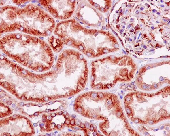
Immunohistochemical staining of paraffin embedded human kidney with purified ab174316 at a working dilution of 1 in 900. The secondary antibody used is a HRP polymer for rabbit IgG. The sample is counter-stained with hematoxylin. Antigen retrieval was perfomed using Tris-EDTA buffer, pH 9.0.

Immunofluorescence staining of Jurkat cells with purified ab174316 at a working dilution of 1 in 300, counter-stained with DAPI. The secondary antibody was Alexa Fluor® 488 goat anti rabbit, used at a dilution of 1 in 200. The cells were fixed in 4% PFA.
Overlay histogram showing Jurkat cells fixed in 2% PFA and stained with purified ab174316 at a dilution of 1 in 90 (pink line). The secondary antibody used was FITC goat anti-rabbit at a dilution of 1 in 150. Rabbit monoclonal IgG was used as an isotype control.
![All lanes : Anti-LOX antibody [EPR4025] (ab174316) at 1/1100 dilution (unpurified)Lane 1 : WI-38 cell lysateLane 2 : Jurkat cell lysateLane 3 : Mouse brainLane 4 : Rat brainLysates/proteins at 20 µg per lane.SecondaryHRP goat anti-rabbit (H+L) at 1/1000 dilution](http://www.bioprodhub.com/system/product_images/ab_products/2/sub_3/17042_ab174316-239763-174316-WB-1.jpg)
All lanes : Anti-LOX antibody [EPR4025] (ab174316) at 1/1100 dilution (unpurified)Lane 1 : WI-38 cell lysateLane 2 : Jurkat cell lysateLane 3 : Mouse brainLane 4 : Rat brainLysates/proteins at 20 µg per lane.SecondaryHRP goat anti-rabbit (H+L) at 1/1000 dilution
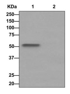
![All lanes : Anti-LOX antibody [EPR4025] (ab174316) at 1/1000 dilution (unpurified)Lane 1 : HeLa cell lysatesLane 2 : Jurkat cell lysatesLane 3 : WI-38 cell lysatesLysates/proteins at 10 µg per lane.](http://www.bioprodhub.com/system/product_images/ab_products/2/sub_3/17043_ab174316-199251-ab1743161.jpg)
All lanes : Anti-LOX antibody [EPR4025] (ab174316) at 1/1000 dilution (unpurified)Lane 1 : HeLa cell lysatesLane 2 : Jurkat cell lysatesLane 3 : WI-38 cell lysatesLysates/proteins at 10 µg per lane.
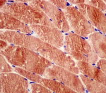
Immunohistochemical analysis of paraffin-embedded human muscle tissue labeling LOX with unpurified ab174316 at a 1/50 dilution.
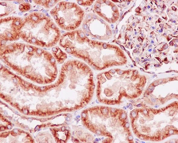
Immunohistochemical staining of paraffin embedded human kidney with unpurified ab174316 at a working dilution of 1 in 300. The secondary antibody used is a HRP polymer for rabbit IgG. The sample is counter-stained with hematoxylin. Antigen retrieval was perfomed using Tris-EDTA buffer, pH 9.0.
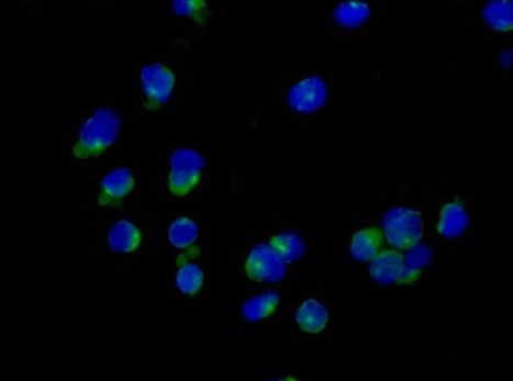
Immunofluorescence staining of Jurkat cells with unpurified ab174316 at a working dilution of 1 in 100, counter-stained with DAPI. The secondary antibody was Alexa Fluor® 488 goat anti rabbit, used at a dilution of 1 in 200. The cells were fixed in 4% PFA.
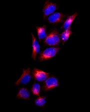
Immunofluorescence analysis of HeLa cells labeling LOX with unpurified ab174316 at a 1/50 dilution.
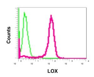
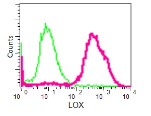
Flow cytometric analysis of permeabilized Jurkat cells using unpurified ab174316 at a 1/10 dilution (red) or a rabbit IgG (negative) (green).
Western blot analysis on immunoprecipitation pellet from (Lane 1) WI-38 cells lysate or (Lane 2) 1X PBS (negative control) using unpurified ab174316 at a 1/10 dilution and HRP-conjugated anti-rabbit IgG preferentially detecting the non-reduced form of rabbit IgG.