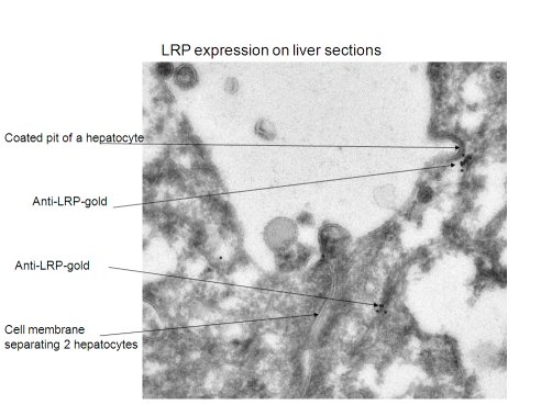
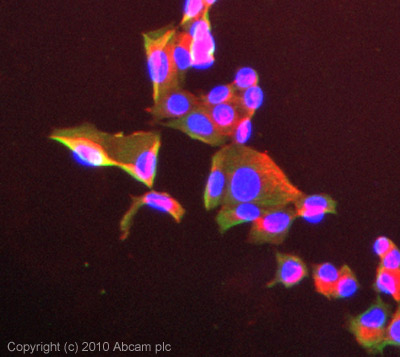
Anti-LRP1 antibody [8G1]
| Name | Anti-LRP1 antibody [8G1] |
|---|---|
| Supplier | Abcam |
| Catalog | ab20384 |
| Prices | $390.00 |
| Sizes | 200 µg |
| Host | Mouse |
| Clonality | Monoclonal |
| Isotype | IgG1 |
| Clone | 8G1 |
| Applications | IHC-F IHC-P WB ICC/IF ICC/IF Electron microscopy DB FC |
| Species Reactivities | Human |
| Antigen | Full length native protein (purified) corresponding to LRP1 aa 1-172 |
| Description | Mouse Monoclonal |
| Gene | LRP1 |
| Conjugate | Unconjugated |
| Supplier Page | Shop |
Product images
Product References
High molecular mass assemblies of amyloid-beta oligomers bind prion protein in - High molecular mass assemblies of amyloid-beta oligomers bind prion protein in
Dohler F, Sepulveda-Falla D, Krasemann S, Altmeppen H, Schluter H, Hildebrand D, Zerr I, Matschke J, Glatzel M. Brain. 2014 Mar;137(Pt 3):873-86.
Differential regulation of extracellular tissue inhibitor of metalloproteinases-3 - Differential regulation of extracellular tissue inhibitor of metalloproteinases-3
Scilabra SD, Troeberg L, Yamamoto K, Emonard H, Thogersen I, Enghild JJ, Strickland DK, Nagase H. J Biol Chem. 2013 Jan 4;288(1):332-42.
A pilot study on low-density lipoprotein receptor-related protein-1 in Chinese - A pilot study on low-density lipoprotein receptor-related protein-1 in Chinese
Chan CY, Chan YC, Cheuk BL, Cheng SW. Eur J Vasc Endovasc Surg. 2013 Nov;46(5):549-56.
.
Sequence identity between the alpha 2-macroglobulin receptor and low density - Sequence identity between the alpha 2-macroglobulin receptor and low density
Strickland DK, Ashcom JD, Williams S, Burgess WH, Migliorini M, Argraves WS. J Biol Chem. 1990 Oct 15;265(29):17401-4.


