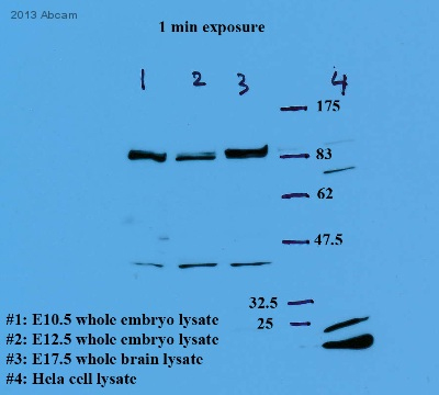
All lanes : Anti-Mad2L2 antibody (ab115622) at 1/1000 dilutionLane 1 : Mouse E10.5 whole embryo lysateLane 2 : Mouse E12.5 whole embryo lysateLane 3 : Mouse E17.5 whole brain lysateLane 4 : HeLa cell lysateSecondaryHRP-conjugated Goat anti-rabbit IgG at 1/5000 dilutiondeveloped using the ECL techniquePerformed under reducing conditions.

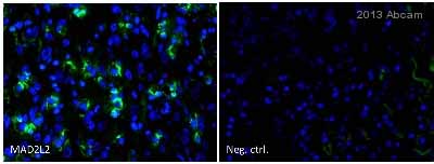
ab115622 staining Mad2L2 (green) in Human lung cancer cells by ICC/IF (Immunocytochemistry/immunofluorescence). Cells were fixed with the Hope technique. Samples were incubated with primary antibody (1/300) for 30 minutes at 25°C. An Alexa Fluor®488-conjugated Goat anti-rabbit IgG polyclonal (1/200) was used as the secondary antibody. Right - negative control.See Abreview

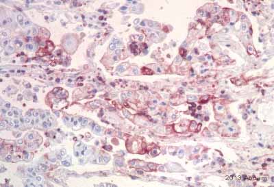
ab115622 staining Mad2L2 in Human lung cancer tissue sections by Immunohistochemistry (IHC-P - paraformaldehyde-fixed, paraffin-embedded sections). Tissue was fixed with the Hope Technique and blocked for 15 minutes at 25°C. Samples were incubated with primary antibody (1/300) for 1 hour at 25°C. An undiluted HRP-conjugated Goat anti-rabbit IgG polyclonal was used as the secondary antibody.See Abreview

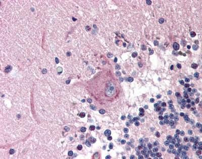
ab115622, at 10µg/ml, staining Mad2L2 in Formalin-fixed, Paraffin-embedded Human Brain Cerebellum tissue by Immunohistochemistry followed by biotinylated secondary antibody, alkaline phosphatase-streptavidin and chromogen.


