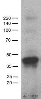
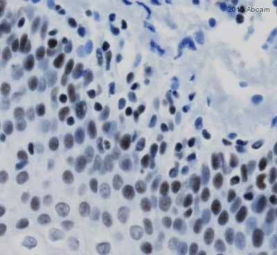
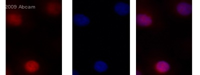
Anti-MBD2 antibody
| Name | Anti-MBD2 antibody |
|---|---|
| Supplier | Abcam |
| Catalog | ab38646 |
| Prices | $395.00 |
| Sizes | 400 µl |
| Host | Rabbit |
| Clonality | Polyclonal |
| Isotype | IgG |
| Applications | ICC/IF ICC/IF IP ELISA WB IHC-P |
| Species Reactivities | Mouse, Rat, Human |
| Antigen | A KLH conjugated synthetic peptide (10-30 aa in length) in the region of 160-177 of MBD2 (Human) |
| Description | Rabbit Polyclonal |
| Gene | MBD2 |
| Conjugate | Unconjugated |
| Supplier Page | Shop |
Product images
Product References
Hiwi mediated tumorigenesis is associated with DNA hypermethylation. - Hiwi mediated tumorigenesis is associated with DNA hypermethylation.
Siddiqi S, Terry M, Matushansky I. PLoS One. 2012;7(3):e33711.
UV radiation regulates Mi-2 through protein translation and stability. - UV radiation regulates Mi-2 through protein translation and stability.
Burd CJ, Kinyamu HK, Miller FW, Archer TK. J Biol Chem. 2008 Dec 12;283(50):34976-82.


