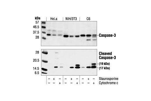
Western blot analysis of extracts from HeLa, NIH/3T3 and C6 cells untreated, staurosporine-treated (3hrs, 1 µM in vivo) or cytochrome c-treated (1hr, 0.25 mg/ml in vitro), using Caspase-3 Antibody #9662 (upper) or Cleaved Caspase-3 (Asp175) Antibody (lower).
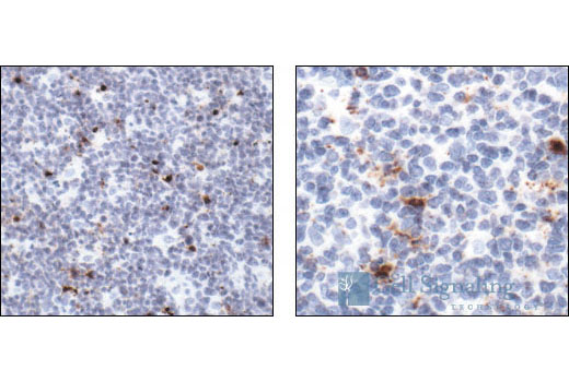
Immunohistochemical analysis of paraffin-embedded human tonsil, showing cytoplasmic and perinuclear localization in apoptotic cells (low and high magnification), using Cleaved Caspase-3 (Asp175) Antibody.
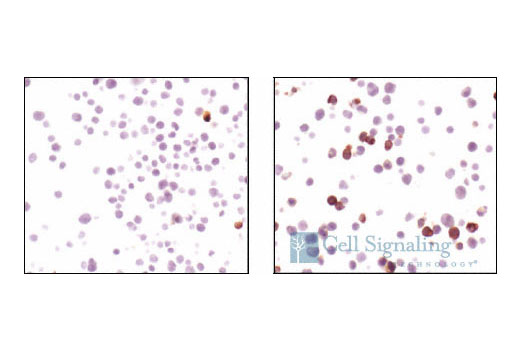
Immunohistochemical analysis using Cleaved caspase-3 (Asp175) antibody on SignalSlide™ Cleaved Caspase-3 IHC controls #8104 (paraffin-embedded Jurkat cells, untreated (left) or etoposide-treated (right)).
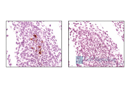
Immunohistochemical analysis of paraffin-embedded mouse embryo, using Cleaved Caspase-3 (Asp175) Antibody preincubated with control peptide (left) or Cleaved Caspase-3 (Asp175) Blocking Peptide #1050 (right).
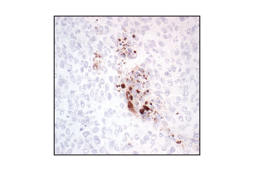
Immunohistochemical analysis of frozen H1650 xenograft section, using Cleaved Caspase-3 (Asp175) Antibody.
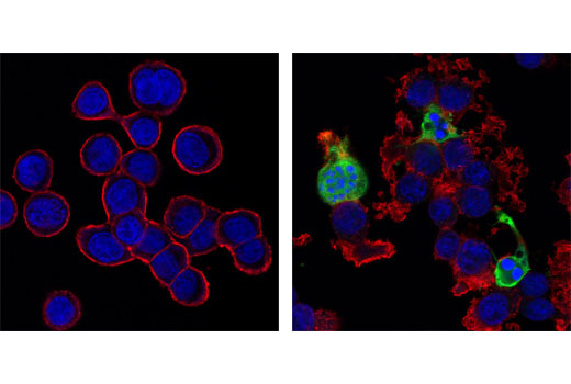
Confocal immunofluorescent images of HT-29 cells, untreated (left) or Staurosporine #9953 treated (right), labeled with Cleaved Caspase-3 (Asp175) Antibody (green). Actin filaments have been labeled with Alexa Fluor ® 555 phalloidin #8953 (red). Blue pseudocolor = DRAQ5 ® (fluorescent DNA dye).
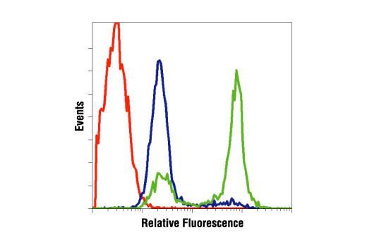
Flow cytometric analysis of Jurkat cells, untreated (blue) or treated with etoposide #2200 (green), using Cleaved Caspase-3 (Asp175) Antibody compared to a nonspecific negative control antibody (red).


