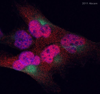
ab69629 staining MGMT in T98G human glioma cell line by Immunocytochemistry/ Immunofluorescence. Cells were fixed in paraformaldehyde and permeabilized in 0.1% Triton X-100 prior to blocking in 3% BSA for 30 minutes at 20°C. The primary antibody was diluted 1/100 and incubated with the sample for 1 hour at 20°C. The secondary antibody was Alexa Fluor® 555-conjugated goat anti-rabbit polyclonal, diluted 1/300. MGMT staining is shown in red (AF 555) and stains nuclei and to a small extent, cytoplasm. Nuclei were stained with DAPI (blue), while microtubules were stained using an anti-tubulin FITC conjugate (green).See Abreview

